| You know you have a skeleton, but did you know that your cells have skeletons, too? Cellular skeletons, or cytoskeletons, are shapeshifting networks of tiny protein filaments, enabling cells to propel themselves, carry cargo, and divide. | |
| Now, an interdisciplinary team of Caltech researchers has designed a way to study and manipulate the cytoskeleton in test tubes in the lab. Understanding how cells control movement could one day lead to tiny, bioinspired robots for therapeutic applications. The work also contributes to the development of new tools for manipulating fluids on very small scales relevant to molecular biology and chemistry. | |
| The work is described in a paper appearing in the journal Nature (“Controlling organization and forces in active matter through optically defined boundaries”). | |
| The building blocks of the cellular cytoskeleton are thin, tube-like filaments called microtubules that can form together into three-dimensional scaffolds. Each microtubule is 1,000 times thinner than a human hair and only about 10 micrometers long (about 1,000 times smaller than a common black ant). Along with motor proteins that power movement, these incredibly small structures combine to propel the relatively large cell–like ants steering and powering a car. |
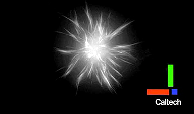
| In previous studies, researchers have taken these molecules out of the cell and put them into test tubes, where the tubules and motor proteins spontaneously group together to organize themselves into star-shaped structures called asters. How asters in a test tube are related to a cytoskeleton powering cell movement, however, is still unclear. Moreover, the collective microtubule organization demonstrated by aster formation involves interacting forces that are not entirely understood. | |
| “What we wanted to know was: how do you get from these spontaneously forming aster structures in the lab, to a cell controlling its movement? And, how can we control these molecules the way a cell does?” says graduate student Tyler Ross, first author on the study. | |
| Led by Ross, a team of Caltech researchers explored how to manipulate the component filaments and motor proteins outside of the cell’s natural environment. In test tubes, they linked motor proteins to light-activated proteins that are naturally found in plants, so that the tubules would only organize into asters when light was shining on them. In this way, the researchers could control when and where asters would form by projecting different patterns of light, enabling them to develop theories about the physical mechanisms underlying aster formation. | |
| Controlling the asters not only allowed for the study of their formation but also enabled the team to build things out of the structures. Ross developed simple procedures of light patterns to place, move, and merge asters of various sizes. The technique offers a way to manipulate structures and study fluid dynamics at a miniscule length scale that is usually difficult to work at; fluids exhibit tricky behaviors at such small volumes. | |
| “Generally, it’s really difficult to manipulate fluids and structures on this length scale. But this is the scale that we’re most interested in for studying cells and chemistry; all of molecular biology works on this scale,” says Ross. “Our light-based system allows us to dynamically manipulate our system. We could look through a microscope and say, ‘Okay we have enough over here, let’s start routing things over there,’ and change the light pattern accordingly. We could use aster structures in such a way that they could stir and mix solutions at very small length scales.” |
Image Credit: Caltech
News This Week
3D-printed implant offers a potential new route to repair spinal cord injuries
A research team at RCSI University of Medicine and Health Sciences has developed a 3-D printed implant to deliver electrical stimulation to injured areas of the spinal cord, offering a potential new route to [...]
Nanocrystals Carrying Radioisotopes Offer New Hope for Cancer Treatment
The Science Scientists have developed tiny nanocrystal particles made up of isotopes of the elements lanthanum, vanadium, and oxygen for use in treating cancer. These crystals are smaller than many microbes and can carry isotopes of [...]
New Once-a-Week Shot Promises Life-Changing Relief for Parkinson’s Patients
A once-a-week shot from Australian scientists could spare people with Parkinson’s the grind of taking pills several times a day. The tiny, biodegradable gel sits under the skin and releases steady doses of two [...]
Weekly injectable drug offers hope for Parkinson’s patients
A new weekly injectable drug could transform the lives of more than eight million people living with Parkinson's disease, potentially replacing the need for multiple daily tablets. Scientists from the University of South Australia [...]
Most Plastic in the Ocean Is Invisible—And Deadly
Nanoplastics—particles smaller than a human hair—can pass through cell walls and enter the food web. New research suggest 27 million metric tons of nanoplastics are spread across just the top layer of the North [...]
Repurposed drugs could calm the immune system’s response to nanomedicine
An international study led by researchers at the University of Colorado Anschutz Medical Campus has identified a promising strategy to enhance the safety of nanomedicines, advanced therapies often used in cancer and vaccine treatments, [...]
Nano-Enhanced Hydrogel Strategies for Cartilage Repair
A recent article in Engineering describes the development of a protein-based nanocomposite hydrogel designed to deliver two therapeutic agents—dexamethasone (Dex) and kartogenin (KGN)—to support cartilage repair. The hydrogel is engineered to modulate immune responses and promote [...]
New Cancer Drug Blocks Tumors Without Debilitating Side Effects
A new drug targets RAS-PI3Kα pathways without harmful side effects. It was developed using high-performance computing and AI. A new cancer drug candidate, developed through a collaboration between Lawrence Livermore National Laboratory (LLNL), BridgeBio Oncology [...]
Scientists Are Pretty Close to Replicating the First Thing That Ever Lived
For 400 million years, a leading hypothesis claims, Earth was an “RNA World,” meaning that life must’ve first replicated from RNA before the arrival of proteins and DNA. Unfortunately, scientists have failed to find [...]
Why ‘Peniaphobia’ Is Exploding Among Young People (And Why We Should Be Concerned)
An insidious illness is taking hold among a growing proportion of young people. Little known to the general public, peniaphobia—the fear of becoming poor—is gaining ground among teens and young adults. Discover the causes [...]
Team finds flawed data in recent study relevant to coronavirus antiviral development
The COVID pandemic illustrated how urgently we need antiviral medications capable of treating coronavirus infections. To aid this effort, researchers quickly homed in on part of SARS-CoV-2's molecular structure known as the NiRAN domain—an [...]
Drug-Coated Neural Implants Reduce Immune Rejection
Summary: A new study shows that coating neural prosthetic implants with the anti-inflammatory drug dexamethasone helps reduce the body’s immune response and scar tissue formation. This strategy enhances the long-term performance and stability of electrodes [...]
Scientists discover cancer-fighting bacteria that ‘soak up’ forever chemicals in the body
A family of healthy bacteria may help 'soak up' toxic forever chemicals in the body, warding off their cancerous effects. Forever chemicals, also known as PFAS (per- and polyfluoroalkyl substances), are toxic chemicals that [...]
Johns Hopkins Researchers Uncover a New Way To Kill Cancer Cells
A new study reveals that blocking ribosomal RNA production rewires cancer cell behavior and could help treat genetically unstable tumors. Researchers at the Johns Hopkins Kimmel Cancer Center and the Department of Radiation Oncology and Molecular [...]
AI matches doctors in mapping lung tumors for radiation therapy
In radiation therapy, precision can save lives. Oncologists must carefully map the size and location of a tumor before delivering high-dose radiation to destroy cancer cells while sparing healthy tissue. But this process, called [...]
Scientists Finally “See” Key Protein That Controls Inflammation
Researchers used advanced microscopy to uncover important protein structures. For the first time, two important protein structures in the human body are being visualized, thanks in part to cutting-edge technology at the University of [...]
