Longer-Term Projects

Vascular Cartographic Scanning Nanodevice (VCSN)
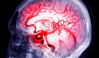
Frank Boehm’s book Nanomedical Device and Systems Design, Challenges, Possibilities, Visions endeavors to explore and present concepts for advanced nanomedical components, devices, and systems that may emerge over the next ~10-30 years. One of these nanodevices is called the Vascular Cartographic Scanning Nanodevice (VCSN) (Figure 1), which would be manifest as an autonomous, ~1 micron in diameter nanomedical device.

Figure 1: Artistic representation of conceptual Vascular Cartographic Scanning Nanodevice (VCSN)
Up to perhaps many thousands, or tens of thousands of VCSN nanodevices might be introduced into the patient intravenously, via ingestion as a pill, through inhalation, transdermally using a patch, or migrate in vivo utilizing a topical gel. Once within the patient their purpose would be to rapidly scan the entire human vasculature down to the smallest capillary lumen (~3 microns in diameter) in ultrahigh-resolution (sub-micron) three-dimensional (3D) digitized format. At all times they would be under the complete control of the surgeon or physician via “outbody” computer commands. Some of the capabilities of the VCSN are listed below:
- Capable of generating a very high-resolution (under 1 micron) 3D rendering of the complete human vasculature down to the smallest capillaries. It may also be applied to the imaging of the lymphatic system, and in a simplified form, the gastrointestinal tract, using a Gastrointestinal Micro Scanning Device (GMSD), (see description below).
- Ability to distinguish vascular and neurological plaque deposits and lesions with high accuracy.
- Capacity to determine vascular wall thicknesses, along with the identification and highlighting of any “hot spot” sites within the vasculature, such as imminent blockages or aneurysms that are at risk of rupturing.
- Surgeon and physicians would be able to “fly-through” all scanned areas via a joystick and computer display for the highly detailed inspection of any desired site within the system. The acquired spatial data from the VCSN may also enable holographic rendering and virtual travel through all imaged systems.
- Ability to facilitate the targeting of tumors by revealing sites of angiogenesis in close proximity to tumor growth sites.
A number of advantages of the VCSN include:
- High compactness and portability as its operation will require a relatively small footprint. This would enable a simple and quick setup and power-up procedures, which will be a boon for applications in developing countries and remote terrestrial environments.In aerospace, it might be utilized as an element of an on board medical diagnostics suite on military and medical aircraft.
- For space travel, it may be reconfigured for integration into spacesuits and spacecraft, and provide a compact yet very powerful medical imaging capability for future Moon and Mars habitats.
- Frugal energy consumption.
- Inexpensive administration and operation
- Rapid scanning time (~5 minutes).
- Ultrahigh resolution digital imagery and inherent flexibility for display across several formats and ease of file transmission to medical personnel globally via secure telecommunications connectivity
- Potential for enabling the significant reduction or elimination of long waiting queues for critical imaging technologies.
Gastrointestinal Micro Scanning Device (GMSD)
The Gastrointestinal Micro Scanning Device (GMSD) (Figure 2) will serve as far less complex precursor to the VCSN as it will not have the capacity for propulsion or navigation. It might, however, employ nascent forms of quantum computing, nanoelectronics, spatial data acquisition, and Pixel Matrix (see below) technologies that are envisaged for the VCSN. Hence, in addition to serving as formative in vivo spatial data acquisition device, the GMSD may also have utility as a test bed of sorts that is employed to identify and resolve technical, integrative, and functional issues toward the development of the VCSN.

![]()
Figure 2: Artistic representation of conceptual Gastrointestinal Micro Scanning Device (GMSD)
The GMSD system will consist of three distinct components that work in unison to generate very high-resolution 3D topography of the entire internal surface of the GIT. The GMSD would accomplish this task by utilizing:
- Bright Ball (BB) scanning device, which would have a smooth spherical morphology of ~3 mm in diameter
- Pulse Generator/Data Transfer (PGDT) unit would trigger the activity of the internalized BB device via a specifically encrypted signal. The BB would be activated to scan subsequently to receiving this specific signal only. Once activated, the BB would commence to transmit a constant spatial data stream to the PGDT, which would serve as a data transfer device when linked to a computer and the Pixel Matrix (see below) software. The PGDT would be securely affixed to the patient’s abdominal surface and would stay in place for the duration of the scan.
- The Pixel Matrix display would process the PGDT supplied spatial data toward the reconstruction a high resolution “pixel per hit” 3D rendering of the total scanned area of the GIT that has been traversed by the BB. This software would enable “fly-through” and cross-sectional capabilities, allowing medical personnel to traverse the entire GIT (using a joystick, computer mouse, or touch screen display) to investigate any potential problem areas in great detail. The spatial data might also be converted to holographic and virtual reality formats. Using this procedure, the physician and/or surgeon would thus recognize any anomalous topography that is associated with tumor growth, lesions, and other abnormal features that may exist within the GIT.
The operational procedure for the GMSD would be relatively simple to implement, as the BB would be introduced orally to the patient in the same manner as a pill. Subsequently, an adhesive waterproof thin film PGDT patch would be affixed to the skin of patient’s abdomen. At this juncture, a system calibration would be performed to ensure that the communication link between the BB and the PGDT is functioning properly. An initial test scan would also be conducted in order to configure the image resolution. The PGDT would emit a unique pulsed signal (e.g., ultrasonic, near-infrared), which when received by sensors embedded within the surface of the BB would trigger all of the embedded emitters/receivers to fire and emit their scanning beams simultaneously in every direction. Once these procedures are completed the patient would be allowed to leave the physician’s office, clinic, or hospital to go about his/her normal routine. The internalized BB would now move along with the natural peristaltic rhythms of the GIT and be naturally eliminated at the conclusion of the transit duration. The patient would then return to the facility in two or three days (contingent on the assessed GIT transit time) to have the PGDT patch removed.
During the designated scanning period, the PGDT will have been continuously uploading spatial data provided by the BB, which would then be interfaced with a computer via a USB port to stream all of this data to the PM software housed within the computer. The data would now be translated to high-resolution 3D imagery on a display. The PM software would calculate BB orientation and would correlate the interrogating hits obtained within predetermined parameters to construct a cross section of the GIT to depict its internal topography. These digitized fragments would then be sequentially pieced together to form a seamless spatially accurate rendering of the system.
News
Electrostatic Discharge Boosts Triboelectric Nanogenerator Current and Enables DC Output
Controlled electrical discharges could enable triboelectric nanogenerators to achieve higher peak currents, extending nano-enabled energy harvesting into chemical processing and self-powered sensing. Paper: Electrostatic discharge as a breakthrough strategy for triboelectric nanogenerators. A new review [...]
Swiss laboratory uses old drugs against rare diseases
Researchers at the University of Geneva are combing through collections of approved drugs to find new therapies for rare diseases – with some success. This approach is gaining traction around the world, while pharmaceutical [...]

Nanozyme Aptasensors Show Promise for Faster Food, Health, and Environmental Testing
By pairing robust artificial enzymes with highly selective aptamers, nanozyme aptasensors could help detect disease biomarkers, pathogens, and contaminants faster, but the review shows that real-world deployment still depends on overcoming matrix interference, biofouling, [...]

Paralyzed Man Feels Sensation Again With Brain Stimulation Device
Aneuroprosthetic system has allowed a man with paralysis to grasp and lift objects and feel touch again. The device helped 42-year-old Keith Thomas of Massapequa, New York, who was paralyzed from the chest down [...]
Global Cancer Cases Could Surge 67% by 2050, New Report Warns
New data reveal major geographic disparities and highlight the urgent need for global action on prevention, early detection, and equitable access to treatment. For roughly one in five people worldwide, cancer will become part [...]

A Deadly Ebola-Like Virus Is Spreading. Are We Ready?
BU virologist Nancy Sullivan says the Bundibugyo outbreak in the Democratic Republic of the Congo underscores the need for broader outbreak preparedness. The death of a nurse marked the moment health officials recognized that [...]

Why Most Animal Viruses Never Become Human Pandemics
From receptor mismatch to risky human-animal interfaces, this article explains why spillover is common but true pandemic emergence remains rare. Introduction Humans are constantly exposed to animal viruses through farming, wildlife contact, and the [...]

Stem cell organoids repair heart microvessels in coronary artery disease models
A Stanford University team has shown that vascular organoids derived from human stem cells can repair the heart’s microvessel network in pigs with ischaemic heart disease – a proof-of-concept advancement that could open new therapeutic [...]

Goodbye GP waiting rooms, hello prevention at home
Prevention is suddenly everywhere in NHS reform. The recent £340m community pharmacy deal is moving more services onto the high street. Community Diagnostic Centres are being expanded, and the Neighbourhood Health Framework sets out [...]
Ebola control is weakened by mistrust and cultural insensitivity
Effective response depends on cooperation with communities and frontline workers, writes Zaeem ul Haq The current Bundibugyo Ebola outbreak in the Democratic Republic of the Congo (DRC) and Uganda is exposing dangerous gaps in [...]

Building the Brain Requires Millions of Dangerous DNA Breaks
Scientists discovered that building a healthy brain involves an unexpected step: young neurons routinely break and rapidly repair their own DNA. As the brain develops, newly formed nerve cells must travel through tightly packed tissue [...]

One Tiny Change May Explain How Viruses Jump From Bats to Humans
Scientists found that one tiny genetic change may determine whether a bat virus stays in bats or becomes a human threat. Most infectious disease outbreaks begin when a virus or other pathogen crosses from animals into [...]
Scientists Discover 250+ Genes That Could Lead to New Ways To Prevent Melanoma
The world’s largest study of mole genetics identified hundreds of genes tied to melanoma risk, uncovering potential new drug targets and paving the way for more accurate melanoma screening and prevention. Researchers at QIMR [...]
Breakthrough Diabetes Treatment Reprograms the Immune System
An engineered stem cell therapy reversed new-onset Type 1 diabetes in mice by shifting the immune system away from attacking insulin-producing cells. For more than a century, people with Type 1 diabetes have relied [...]
Taking the world’s temperature: WHO chief spotlights global health emergencies
Taking the world’s temperature on pressing health matters, WHO Director-General Tedros Adhanom Ghebreyesus provided the latest on current global challenges - and successes when it comes to international cooperation. “The outbreaks of hantavirus, Ebola and Marburg all show [...]
Scientists Create Tiny “Mini Livers” That Could One Day Replace Liver Transplants
Engineered tissue grafts could help perform key liver functions and benefit thousands of people living with liver failure. The liver is one of the body’s hardest-working organs, carrying out hundreds of vital jobs, from [...]

NanoMedical Brain/Cloud Interface – Explorations and Implications. A new book from Frank Boehm
New book from Frank Boehm, NanoappsMedical Inc Founder: This book explores the future hypothetical possibility that the cerebral cortex of the human brain might be seamlessly, safely, and securely connected with the Cloud via [...]
Scientists Discover Surprising Way To Help the Brain Recover After Stroke
A new study suggests that strengthening the body’s natural circadian rhythms may help the brain recover after stroke, even when treatment begins days after the injury. Every year, millions of people survive a stroke, [...]

Our books now available worldwide!
Online Sellers other than Amazon, Routledge, and IOPP Indigo Global Health Care Equivalency in the Age of Nanotechnology, Nanomedicine and Artifcial Intelligence Global Health Care Equivalency In The Age Of Nanotechnology, Nanomedicine And Artificial [...]

Younger Generations Are Aging Faster – and It May Be Fueling a Surge in Cancer
Younger generations may be aging biologically faster than those before them, and that shift could help explain rising rates of cancer at younger ages. For decades, cancer was viewed largely as a disease of [...]