Modern light microscopic techniques provide extremely detailed insights into organs, but the terabytes of data they produce are usually nearly impossible to process. New software, developed by a team led by MDC scientist Dr. Stephan Preibisch and presented in Nature Methods (“BigStitcher: Reconstructing high-resolution image datasets of cleared and expanded samples”), is helping researchers make sense of these reams of data.
It works almost like a magic wand. With the help of a few chemical tricks and ruses, scientists have for a few years now been able to render large structures like mouse brains and human organoids transparent. CLARITY is perhaps the most well-known of the many different sample clearing techniques, with which almost any object of study can be made nearly as transparent as water. This enables researchers to investigate cellular structures in ways they could previously only dream of.
And that’s not all. In 2015 another conjuring trick – called expansion microscopy – was presented in the journal Science. A research team at Massachusetts Institute of Technology (MIT) in Cambridge discovered that it was possible to expand ultrathin slices of mouse brains nearly five times their original volume, thereby allowing samples to be examined in even greater detail.
The software brings orders to the data chaos
“With the aid of modern light-sheet microscopes, which are now found in many labs, large samples processed by these methods can be rapidly imaged,” says Dr. Stephan Preibisch, head of the MDC research group on Microscopy, Image Analysis & Modeling of Developing Organisms. “The problem, however, is that the procedure generates such large quantities of data – several terabytes – that researchers often struggle to sift through and organize the data.”
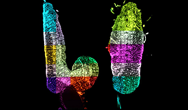
To create order in the chaos, Preibisch and his team have now developed a software program that after complex reconstructing the data resembles somewhat Google Maps in 3D mode. “One can not only get an overview of the big picture, but can also zoom in to specifically examine individual structures at the desired resolution,” explains Preibisch, who has christened the software “BigStitcher.” Now, the computer program, which any interested scientist can use, has been presented in the scientific journal Nature Methods.
A team of twelve researchers from Berlin, Munich, the United Kingdom, and the United States were involved in the development. The paper’s two lead authors are David Hörl, from Ludwig-Maximilians-Universität München and the Berlin Institute for Medical Systems Biology (BIMSB) of the MDC, as well as Dr. Fabio Rojas Rusak from the MDC research group of Professor Mathias Treier. The researchers show in their paper that algorithms can be used to reconstruct and scale the data acquired by light-sheet microscopy in such a way that renders a supercomputer unnecessary. “Our software runs on any standard computer,” says Preibisch. “This allows the data to be easily shared across research teams.“
Image Credit: Janelia / MDC
News This Week
Electrostatic Discharge Boosts Triboelectric Nanogenerator Current and Enables DC Output
Controlled electrical discharges could enable triboelectric nanogenerators to achieve higher peak currents, extending nano-enabled energy harvesting into chemical processing and self-powered sensing. Paper: Electrostatic discharge as a breakthrough strategy for triboelectric nanogenerators. A new review [...]
Swiss laboratory uses old drugs against rare diseases
Researchers at the University of Geneva are combing through collections of approved drugs to find new therapies for rare diseases – with some success. This approach is gaining traction around the world, while pharmaceutical [...]
Nanozyme Aptasensors Show Promise for Faster Food, Health, and Environmental Testing
By pairing robust artificial enzymes with highly selective aptamers, nanozyme aptasensors could help detect disease biomarkers, pathogens, and contaminants faster, but the review shows that real-world deployment still depends on overcoming matrix interference, biofouling, [...]

Paralyzed Man Feels Sensation Again With Brain Stimulation Device
Aneuroprosthetic system has allowed a man with paralysis to grasp and lift objects and feel touch again. The device helped 42-year-old Keith Thomas of Massapequa, New York, who was paralyzed from the chest down [...]
Global Cancer Cases Could Surge 67% by 2050, New Report Warns
New data reveal major geographic disparities and highlight the urgent need for global action on prevention, early detection, and equitable access to treatment. For roughly one in five people worldwide, cancer will become part [...]
A Deadly Ebola-Like Virus Is Spreading. Are We Ready?
BU virologist Nancy Sullivan says the Bundibugyo outbreak in the Democratic Republic of the Congo underscores the need for broader outbreak preparedness. The death of a nurse marked the moment health officials recognized that [...]
Why Most Animal Viruses Never Become Human Pandemics
From receptor mismatch to risky human-animal interfaces, this article explains why spillover is common but true pandemic emergence remains rare. Introduction Humans are constantly exposed to animal viruses through farming, wildlife contact, and the [...]

Stem cell organoids repair heart microvessels in coronary artery disease models
A Stanford University team has shown that vascular organoids derived from human stem cells can repair the heart’s microvessel network in pigs with ischaemic heart disease – a proof-of-concept advancement that could open new therapeutic [...]

Goodbye GP waiting rooms, hello prevention at home
Prevention is suddenly everywhere in NHS reform. The recent £340m community pharmacy deal is moving more services onto the high street. Community Diagnostic Centres are being expanded, and the Neighbourhood Health Framework sets out [...]
Ebola control is weakened by mistrust and cultural insensitivity
Effective response depends on cooperation with communities and frontline workers, writes Zaeem ul Haq The current Bundibugyo Ebola outbreak in the Democratic Republic of the Congo (DRC) and Uganda is exposing dangerous gaps in [...]
Building the Brain Requires Millions of Dangerous DNA Breaks
Scientists discovered that building a healthy brain involves an unexpected step: young neurons routinely break and rapidly repair their own DNA. As the brain develops, newly formed nerve cells must travel through tightly packed tissue [...]

One Tiny Change May Explain How Viruses Jump From Bats to Humans
Scientists found that one tiny genetic change may determine whether a bat virus stays in bats or becomes a human threat. Most infectious disease outbreaks begin when a virus or other pathogen crosses from animals into [...]
Scientists Discover 250+ Genes That Could Lead to New Ways To Prevent Melanoma
The world’s largest study of mole genetics identified hundreds of genes tied to melanoma risk, uncovering potential new drug targets and paving the way for more accurate melanoma screening and prevention. Researchers at QIMR [...]
Breakthrough Diabetes Treatment Reprograms the Immune System
An engineered stem cell therapy reversed new-onset Type 1 diabetes in mice by shifting the immune system away from attacking insulin-producing cells. For more than a century, people with Type 1 diabetes have relied [...]
Taking the world’s temperature: WHO chief spotlights global health emergencies
Taking the world’s temperature on pressing health matters, WHO Director-General Tedros Adhanom Ghebreyesus provided the latest on current global challenges - and successes when it comes to international cooperation. “The outbreaks of hantavirus, Ebola and Marburg all show [...]
Scientists Create Tiny “Mini Livers” That Could One Day Replace Liver Transplants
Engineered tissue grafts could help perform key liver functions and benefit thousands of people living with liver failure. The liver is one of the body’s hardest-working organs, carrying out hundreds of vital jobs, from [...]