Over the years, researchers have tried hard to comprehend topographic signals that promote cell mechanical sensitive responses. The extracellular matrix (ECM) provides a complex cellular microenvironment that controls cellular behavior. Nevertheless, only a few functions of these factors are understood, and most remain obscure.
An article published in Advanced Sciences presented a convenient method to demonstrate the curved structure of the ECM network that regulates stem cell mechanotransduction. Here, an ECM-mimicking nanofiber network was prepared using electrospinning technology.
![]()
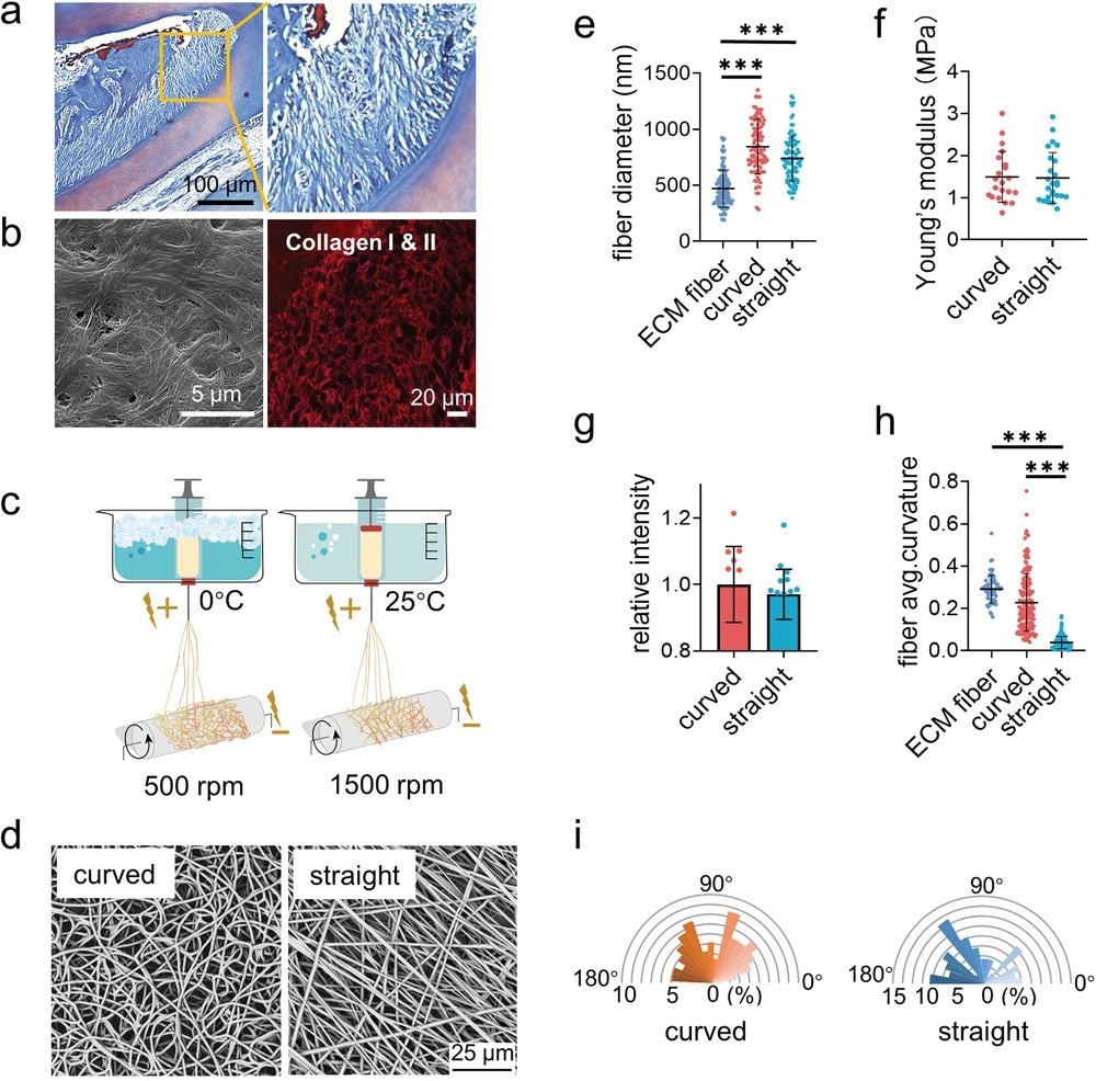
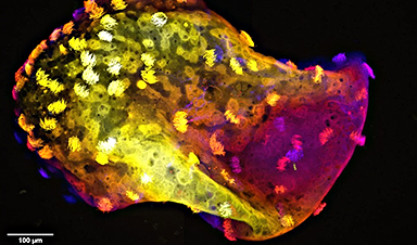
Figure 1. Fabrication and characterization of the curved and straight nanofiber network. a) The Representative images of masson staining of the periodontal tissues. b) The SEM (left) image of the decellularized periodontal ligament tissues and the representative fluorescence image of the collagen I and II (right) in periodontal tissues. c) Scheme of the curved and straight nanofiber network fabrication. The curved and straight fiber network require 0 °C and 25 °C electrospinning temperature, respectively. d) The representative SEM images of the curved and straight fibers (three technical replicates). e) The diameter of the ECM fibers in the periodontal tissues and the artificial fibers (n = 100, two technical replicates). f) Young’s modulus of the curved and straight nanofiber network as detected by Nanoindenter (n = 20, two technical replicates). g) Specific surface area of the curved and straight surfaces as detached by the fluorescent intensity of the adsorbed FITC-BSA at 562 nm (n = 12, two technical replicates). h) The average curvature of the ECM fibers in the periodontal tissues and the artificial fibers (n = 160, two technical replicates). i) The orientation angles (n = 100, two technical replicates) of the curved and straight fibers.
The curved nanofiber promoted cell bridge formation due to cytoskeleton tension. Moreover, the myosin-II-based intracellular force generated by the actomyosin filaments inclined the cell lineage towards osteogenic differentiation. Thus, the present study has provided a better understanding of the effects of topographic signals on cell behavior, thereby aiding the development of new biomaterials.
Effect of Nanofibers on the Functioning of Stem Cells
According to recent studies, the physiological and behavioral functions of cells are influenced by biochemical and physical factors. Novel biomaterials that mimic ECM’s stiffness, degradation, ligand diffusion, stress relaxation, and other physical properties, in addition to the usual chemical effects, have been created.
Nanomaterials, such as nanofibers, are mostly fabricated through electrospinning. In this process, a strong electric field is used to transform solution-based polymers into continuous nanometer-sized fibers.
Various nanofibers differ in their properties, including surface-to-volume ratio and morphology. These characteristics can be altered based on the polymer and intended application. The electrospinning parameters, solution parameters, and ambient characteristics affect the properties of the nanofibers.
Stem cells can develop into various cell types and construct any tissue in the body. However, stem cells have low vitality and are challenging to multiply, which limits their application for a wider range of prospective therapeutic benefits.
Stem cells and electrospun nanofibers have two key advantages. First, by changing the chemical characteristics of the nanofibers to enhance their interactions with stem cells, they can operate as advantageous scaffolds for maintaining stem cells. Second, stem cells can be delivered using nanofibers to particular tissues or organs for tissue engineering and wound repair.
Previous reports have suggested that cancer cells unbend the curled collagen fibers in the ECM during tumor growth. Although curved structures in the fibrous connective tissue, known as the periodontal ligament, were previously known, their function at the cellular level remains unclear. Moreover, studies in this area have been restricted by the absence of techniques for creating curved nanofibers.
Curved Nanofibers to Promote Stem Cell Mechanotransduction
Despite previous reports on electrospinning technology to fabricate biomaterials that mimic the ECM, only a few reports have described the fabrication of curved nanofibers. On the other hand, other studies that carried out low-temperature electrospinning have focused on the porosity of the matrix rather than the topology of nanofibers.
In this study, cryogenic electrospinning technology was utilized to fabricate ECM-mimicking curved nanofibers as a tool to study cell response when exposed to curved structures. Interestingly, curved nanofibers influenced the behavior of stem cells, altering their adhesive nature compared to straight nanofibers.
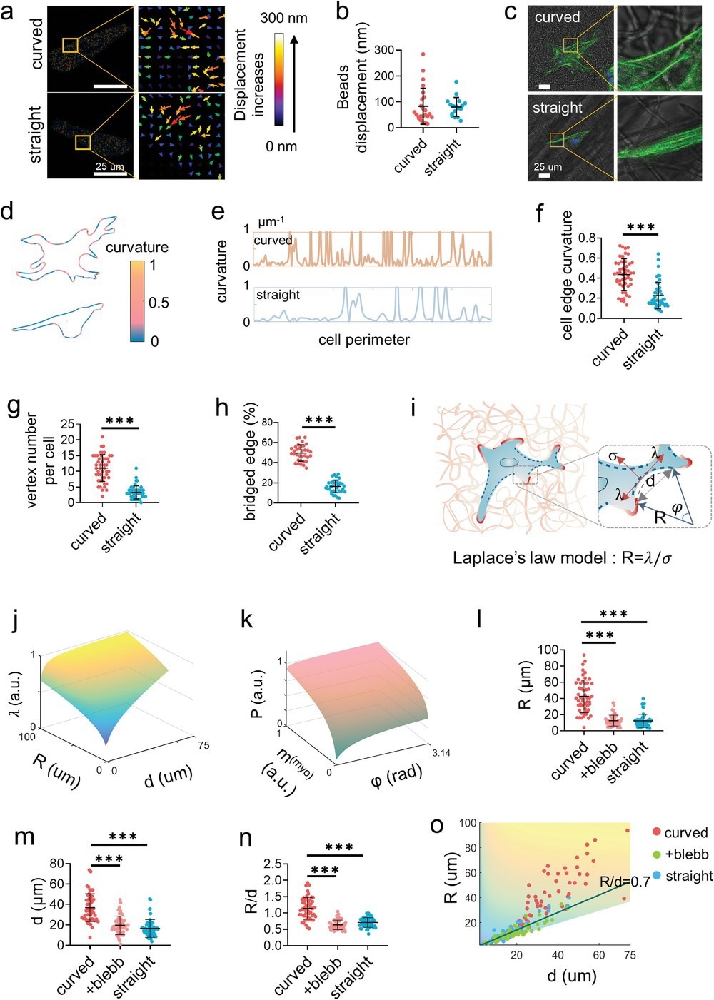
While cells adhered along straight nanofibers, they crossed curved nanofibers to form cell bridges, indicating that the cell bodies overhung instead of attaching to the nanofibers.
The formation of cell bridges rearranged the distribution of the actomyosin cytoskeleton and imparted extra intracellular force, enhancing stem cell mechanotransduction and promoting osteogenic differentiation. The new findings of this study helped obtain a better understanding of the crucial role of biomechanical principles in promoting the development of tissue engineering.
Thus, the present investigation of cell mechanosensing revealed that, while the cell boundary was frequently parallel to the surrounding straight nanofibers, it invariably traversed multiple curved nanofibers as bridges. The cells on the curved nanofibers had a significant percentage of unbound borders that formed large radial arcs that bowed inwards.
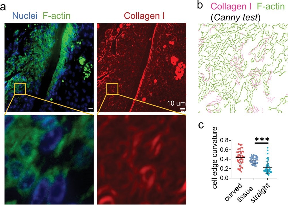
Figure 3. Immunofluorescence staining displays widely distributed cell bridges in the periodontal ligament. a) The representative fluorescence images of nuclei (blue), F-actin (green), and collagen I (red) staining of the mouse periodontal ligament. b) Canny edge test image of the yellow box area in (a). The magenta and green represent the collagen I and F-actin, respectively. c) The average curvature of the cell edges (n = 50, two technical replicates) of the cells in periodontal ligament and cultured on the artificial fibers.
Conclusion
In summary, a simple electrospinning technology that operates at a low speed and temperature to fabricate ECM-mimicking curved nanofiber structures was developed. While the curved nanofibers promoted discrete adhesion in stem cells, straight networks induced the formation of continuous adhesion by stem cells along with the fiber structure.
The curved nanofibers stimulated stem cell mechanotransduction by forming a cell bridge, thereby promoting osteogenic differentiation and proliferation of stem cells. Inducing mechanotransduction and mechanosensing signaling pathways via the formation of nonadhesive bridges caused actomyosin to aggregate and contract.
Thus, the present study demonstrated that the knowledge of cell mechanosensing and tissue development could be improved by using this curved matrix to enhance the database of biomaterials that mimic the ECM.
News
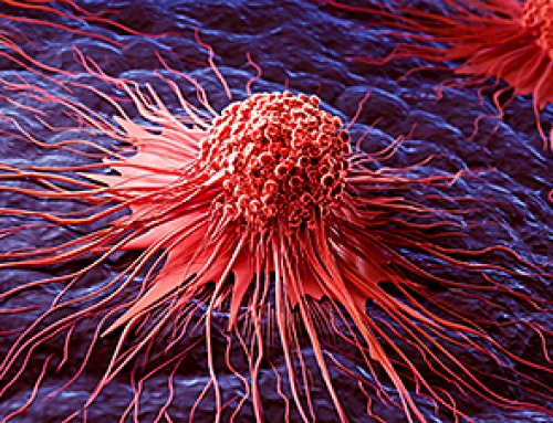
Scientists Uncover Fatal Weakness in “Zombie Cells” Linked to Cancer
A newly identified weakness in “zombie” cells may open the door to more precise cancer treatments by turning their own survival strategy against them. A new class of drugs takes advantage of a recently [...]

Bowel and Ovarian Cancers Are Dramatically Rising in Young Adults, Scientists Aren’t Sure Why
Cancer incidence is increasing, especially among younger adults, and current risk factors don’t fully account for the trend. Scientists suggest other underlying causes may be contributing. Cancer patterns in England are shifting in a [...]
New Immune Pathway Could Supercharge mRNA Cancer Vaccines
A surprising backup system in the immune response to mRNA vaccines may hold the key to more effective cancer treatments. The arrival of mRNA vaccines against SARS-CoV-2 in 2020 marked a turning point in the COVID-19 pandemic. Today, [...]
Scientists Discover “Molecular Switch” That Fuels Alzheimer’s Brain Inflammation
A newly identified trigger of brain inflammation could offer a fresh target for slowing Alzheimer’s progression. The brain has its own built-in immune system that identifies threats and responds to them. In Alzheimer’s disease, growing evidence [...]

Molecular Manufacturing: The Future of Nanomedicine – New book from NanoappsMedical Inc.
This book explores the revolutionary potential of atomically precise manufacturing technologies to transform global healthcare, as well as practically every other sector across society. This forward-thinking volume examines how envisaged Factory@Home systems might enable the cost-effective [...]
Forgotten Medicinal Plant Shows Promise in Fighting Dangerous Superbugs
A traditional medicinal plant, tormentil, shows promise against antibiotic-resistant bacteria in laboratory tests. Its compounds work by limiting bacterial growth and boosting antibiotic performance. Before the development of modern antibiotics, plant-based remedies were commonly [...]

NanoMedical Brain/Cloud Interface – Explorations and Implications. A new book from Frank Boehm
New book from Frank Boehm, NanoappsMedical Inc Founder: This book explores the future hypothetical possibility that the cerebral cortex of the human brain might be seamlessly, safely, and securely connected with the Cloud via [...]

New Research Finds Shocking Link Between Chili Peppers and Cancer
If you love spicy food, you are not alone. But scientists are taking a closer look at whether eating a lot of chili peppers could affect your cancer risk. Could your love of spicy [...]

New book from Nanoappsmedical Inc. – Global Health Care Equivalency
A new book by Frank Boehm, NanoappsMedical Inc. Founder. This groundbreaking volume explores the vision of a Global Health Care Equivalency (GHCE) system powered by artificial intelligence and quantum computing technologies, operating on secure [...]
Scientists Create “Neurobots” – Living Machines With Their Own Nervous Systems
Neurobots—xenobots with neurons—show self-organized nervous systems and enhanced behaviors, revealing new insights into how biology builds functional structures. In 2020, researchers at Tufts University developed tiny living structures known as xenobots using frog cells. These microscopic organisms [...]

Our books now available worldwide!
Online Sellers other than Amazon, Routledge, and IOPP Indigo Global Health Care Equivalency in the Age of Nanotechnology, Nanomedicine and Artifcial Intelligence Global Health Care Equivalency In The Age Of Nanotechnology, Nanomedicine And Artificial [...]

Amazonian Chocolate Could Become the Next Superfood, Scientists Say
New research into Amazonian cocoa reveals that its value may extend beyond flavor alone. Chocolate from the Amazon is already known worldwide for its distinctive taste, but new research suggests it may offer even [...]
Nanobody repairs misfolded CFTR inside cells, boosting function in cystic fibrosis
A tiny antibody component could fundamentally transform the treatment of cystic fibrosis: For the first time, researchers have succeeded in developing a so-called nanobody that penetrates directly into human cells and can repair the [...]

20-Year Study Finds Daily Multivitamins Don’t Extend Lifespan
A large, decades-long study of over 390,000 U.S. adults challenges a widespread assumption about daily multivitamins. Multivitamins are a daily habit for millions of Americans, often taken with the expectation that they will extend [...]
Novel Investment Paradigms for Regenerative Healthcare Ecosystems
Introduction The transition toward regenerative healthcare ecosystems—anchored in wellness optimization, disease prevention, eradication strategies, and healthy longevity—necessitates a structural reconfiguration of capital architectures, governance models, and incentive design. Regenerative healthcare, by definition, transcends episodic [...]

What If Consciousness Exists Beyond Your Brain
Scientists still don’t know how consciousness emerges from the brain. New ideas suggest it may not emerge at all, but instead be a basic feature of reality. Is consciousness produced by the brain, or [...]