
A paper recently published in the journal ACS Omega reviewed the current status and potential of X-ray spectroptychography for the characterization of advanced nanomaterials.
What is X-ray Spectroptychography?
X-ray spectroptychography is an emerging technique that represents the ptychographic version of X-ray spectromicroscopy and is used for the chemical microanalysis of nanomaterials such as batteries and catalysts. This technique is based on the existing synchrotron X-ray spectromicroscopy and microscopy techniques with an added ptychography, an algorithmic imaging technique, for image acquisition.
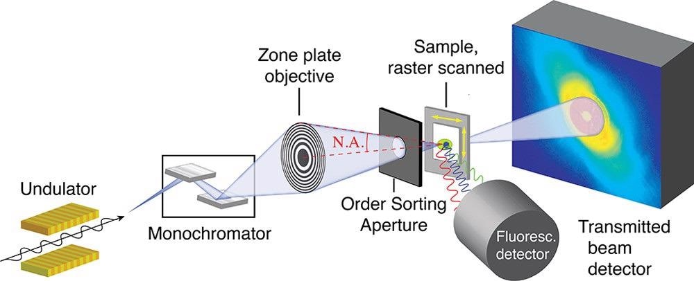
Figure 1. Schematic of a scanning transmission X-ray microscope. Adapted with permission from ref (45). Sample is raster scanned through the focused X-ray beam. In conventional STXM, transmission is measured by a serial detector downstream of the sample. In ptychographic STXM, a diffraction pattern is measured with a pixelated detector downstream of the sample, as shown in the figure. © Urquhart (2022)
X-ray spectroptychography provides a higher spatial resolution during chemical microanalysis compared to conventional X-ray optics. In traditional X-ray microscopes, the spatial resolution is limited by the focusing properties of their X-ray optics. However, ptychography can significantly enhance the spatial resolution of X-ray microscopy as this lensless imaging technique relies on the diffraction of coherent radiation by an amorphous sample.
Within coherent diffractive imaging (CDI), the forerunner of ptychography, the reconstruction of the image of an isolated object is based on the diffraction pattern of the object, which is obtained by the uniform illumination of the object. This factor limits the practical application of the technique. However, unlike the CDI method, ptychography utilizes a sequence of coherent far-field diffraction patterns obtained from the overlapping regions of the isolated sample without requiring the sample.
Ptychography microscopes use zone plates, refractive Laue lenses, Kirkpatrick−Baez mirrors, or pinholes to illuminate the test sample with a well-defined coherent probe. These optics provide a constant focal length when the X-ray energy is altered for spectroscopy.
Both phase and absorption can be utilized for ptychography image contrast. Phase contrast is more crucial in a hard X-ray measurement obtained from a test sample. X-ray spectroptychography can provide the full refractive index of a sample comprising the phase and absorption spectra.
X-Ray Spectroptychography Applications
In the last few years, the spatial resolution provided by X-ray spectroptychography was enhanced owing to the improvements in algorithms and instrumentation associated with the technique. Currently, the technique is used to study nanostructured energy materials such as magnetic materials and fuel cell cathodes.
Magnetic Materials
X-ray magnetic circular dichroism (XMCD) spectroptychography can be used to evaluate the magnetic nanostructure of materials. For instance, XMCD spectroptychography was used to investigate the out-of-plane magnetization in gadolinium/iron multilayer samples and study the nanoscale magnetite single crystals synthesized by magnetotactic bacteria.
Battery Materials
X-ray spectroptychography can provide a chemical characterization of battery materials at a higher spatial resolution compared to conventional X-ray spectromicroscopy and microscopy. Specifically, this technique provides an oxidation state mapping of particular metal ions with a high spatial resolution during battery cycles. For instance, iron L3-edge X-ray spectroptychography was used to map delithiation and lithiation through the corresponding changes in the iron 2p oxidation state.
The metal oxidation state was also mapped at hard X-ray energies using this technique. Spectroptychography is used for chemical mapping to monitor the relationship between the chemical and mechanical stability of lithium-ion batteries. The technique is also used for the elucidation of surface chemistry and minority phases.
Catalyst Materials
X-ray spectroptychography is used extensively to investigate catalyst materials as the improvement of these materials depends on high spatial resolution chemical mapping of heterogeneous materials. Additionally, this technique is also used to evaluate porosity in heterogeneous catalyst materials such as fluid catalytic cracking catalysts.
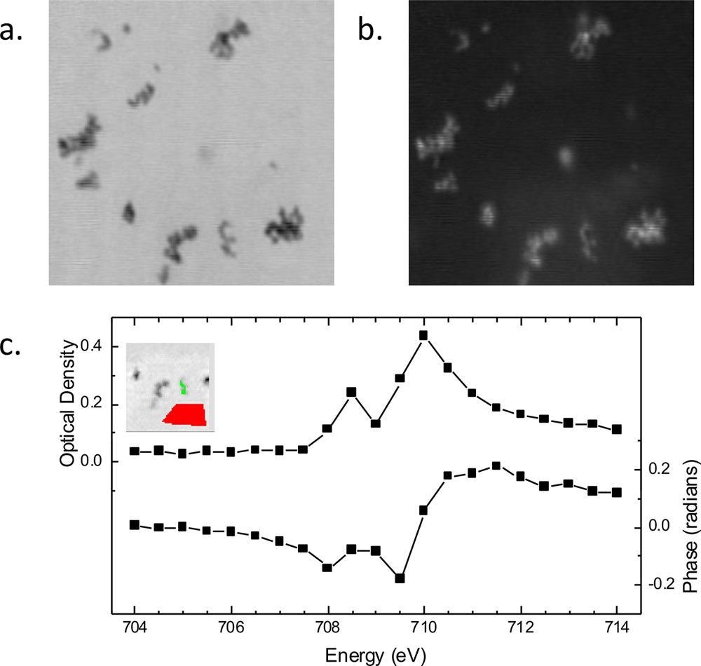
Figure 2. Spectroptychography of 30 nm diameter Fe2O3 nanoparticles. Amplitude (a) and phase (b) ptychography images, recorded at 710.0 eV. Image size is 2.1 × 2.1 μm. (c) Absorption (optical density) and phase Fe L3 spectra obtained from this sample. (Inset) Region from which the sample signal for amplitude and phase (green region) was extracted. Amplitude signal from an open area (red region) was used for the incidence flux in the calculation of the sample optical density using Beer’s law, −ln(I/Io). © Urquhart (2022)
Other Aspects Related to X-ray Spectroptychography
Spectroptychotomography
Three-dimensional (3D) mapping using spectroptychotomography can help in avoiding spurious correlations in two-dimensional (2D) transmission imaging. For instance, a “sparse” spectroptychotomography study based on the simultaneous algebraic reconstruction technique (SART) was used to reduce the number of projections needed to obtain a high-quality reconstruction.
Although multi-energy spectroptychotomography studies can provide chemical mapping in a detailed manner, these studies are often restricted to radiation-resistant materials and are extremely time-consuming.
Extended X-ray Absorption Fine Structure (EXAFS) Spectroptychography
EXAFS spectroptychography requires several photon energies to provide an interferogram, which can be then Fourier transformed to obtain a radial distribution function. However, EXAFS spectroptychography studies require excellent stability and are time-consuming.
In Situ and Operando Spectroptychography
This technique can be potentially used to investigate samples in realistic and varied conditions. However, the dwell times needed for ptychography measurements and the requirement for several energies for spectroscopic sensitivity are the major challenges of in situ studies.
X-ray Linear Dichroism Spectroptychography
This technique can be used in several applications. For instance, the linear dichroism of a vanadium pentoxide crystal was investigated by X-ray linear dichroism spectroptychography at orthogonal polarization states and the V K edge to obtain phase and absorption maps of polycrystalline vanadium pentoxide.
Future of X-ray Spectroptychography
X-ray spectroptychography and ptychography are rapidly becoming the preferred synchrotron techniques for optics and instrumentation. However, the phase spectra and images of materials with complex structures obtained by these techniques cannot be easily rationalized, which is a major drawback. Phase spectra can reveal additional sample-related information, while phase images can be used to a limited extent for chemical imaging of samples with decreased radiation damage.
Additionally, X-ray cameras display reduced sensitivity at lower photon energies, which limits the application of spectroptychography in different materials such as lithium-ion batteries and organic electronic batteries. A more consistent signal-to-noise ratio is required to obtain higher spatial resolution measurements. Recent studies have demonstrated that spectroptychography has significant potential for correlative imaging when it is used with other microscopy techniques.
Complex ptychographic data sets can be analyzed using statistical methods such as machine learning, which can help in the translation of advanced statistical analysis and reconstruction methods to experimentally accessible tools.
To summarize, X-ray spectroptychography is fast becoming a mainstream method for the chemical microanalysis of nanomaterials. However, the time required for data acquisition remains a challenge in this technique.
News

Our books now available worldwide!
Online Sellers other than Amazon, Routledge, and IOPP Indigo Global Health Care Equivalency in the Age of Nanotechnology, Nanomedicine and Artifcial Intelligence Global Health Care Equivalency In The Age Of Nanotechnology, Nanomedicine And Artificial [...]

Younger Generations Are Aging Faster – and It May Be Fueling a Surge in Cancer
Younger generations may be aging biologically faster than those before them, and that shift could help explain rising rates of cancer at younger ages. For decades, cancer was viewed largely as a disease of [...]

Using Cannabis Could Raise Your Stroke Risk by 37%, Massive Study Reveals
Large-scale evidence suggests cannabis, cocaine, and amphetamines may directly raise stroke risk, including in younger adults. As recreational drug use becomes increasingly common, researchers are uncovering evidence that its health consequences may extend far beyond [...]

Could Vitamin C Be the Secret to Keeping Your Brain Younger?
Lower vitamin C levels were linked to reduced brain volume and weaker neural connectivity in older adults, suggesting a potential connection between nutrition and brain health. Could a common vitamin help preserve the brain [...]

This Deadly Disease Was Wiping Out Humans 5,500 Years Ago
A new study suggests plague was already a deadly threat 5,500 years ago, striking small hunter-gatherer communities long before cities and agriculture emerged. For centuries, plague has been remembered as the disease that devastated [...]

China closing in but US leads in biotech quality, commercial reach, survey finds
SAN DIEGO, June 22 (Reuters) - China, which now conducts more clinical drug trials, opens new tab than the U.S., still lags in the quality and commercial reach of its biomedical science, according to a recent survey, opens new [...]

New method generates renewable supply of progenitor immune cells
In a paper published in Cell, a USC Stem Cell-led team reports a new way of generating a renewable and expandable supply of the progenitor cells that give rise to macrophages. These immune cells help [...]

Scientists Just Discovered a Cellular Survival System That Was Never Supposed To Exist
A surprising backup pathway allows cells to make a crucial amino acid when their primary machinery fails. For decades, biologists believed cells had only one way to access a molecule they cannot live without. New [...]

Artificial cells gain porous membranes, enabling lab reactions and drug release
Artificial cells created in the laboratory offer a wide range of potential applications. Until now, however, their membranes—unlike those of real cells—have been virtually impermeable. Researchers at the Max Planck Institute for Polymer Research, [...]

Popular Weight-Loss Drugs Like Ozempic Linked to Lower Breast Cancer Risk
Ozempic and similar weight-loss drugs were linked to a striking 30% reduction in breast cancer risk in a study of more than 110,000 women. Popular weight-loss and diabetes medications such as Ozempic, Wegovy, Mounjaro, [...]

Stanford Scientists Discover Explosive New Type of Immune Cell
Scientists studying the remarkable regenerative abilities of planarian flatworms have uncovered a previously unknown type of immune cell with an unusually destructive defense strategy. What if an immune cell could wipe out nearby threats [...]

Big Pharma-backed SonoThera sounds off with $125M series B for bubble-based genetic delivery
Bay Area biotech SonoThera is bubbling to a clinical boil after raising a $125 million series B with the backing of some of the biggest names in pharma. Vida Ventures led the raise, with the venture [...]

Joint initiative of 5 EU countries calls for ‘unified approach’ to pharma framework amid US drug pricing pressure
With drug pricing pressure building from the U.S., a healthcare-focused consortium of five European countries is calling for a “unified approach” to strengthen Europe’s pharmaceutical framework and access to innovative medicines. Belgium, the Netherlands, [...]

Molecular Manufacturing: The Future of Nanomedicine – New book from NanoappsMedical Inc.
This book explores the revolutionary potential of atomically precise manufacturing technologies to transform global healthcare, as well as practically every other sector across society. This forward-thinking volume examines how envisaged Factory@Home systems might enable the cost-effective [...]

NanoMedical Brain/Cloud Interface – Explorations and Implications. A new book from Frank Boehm
New book from Frank Boehm, NanoappsMedical Inc Founder: This book explores the future hypothetical possibility that the cerebral cortex of the human brain might be seamlessly, safely, and securely connected with the Cloud via [...]

New book from Nanoappsmedical Inc. – Global Health Care Equivalency
A new book by Frank Boehm, NanoappsMedical Inc. Founder. This groundbreaking volume explores the vision of a Global Health Care Equivalency (GHCE) system powered by artificial intelligence and quantum computing technologies, operating on secure [...]