Scientists at EMBL have captured how human chromosomes fold into their signature rod shape during cell division, using a groundbreaking method called LoopTrace.
By observing overlapping DNA loops forming in high resolution, they revealed that large loops form first, followed by nested smaller loops, all repelling each other into compact structures. This new insight not only reshapes our understanding of chromosome mechanics but could also help explain errors that lead to cancer and genetic disorders.
The Mystery of Chromosome Division
One of the most remarkable abilities of living cells is their capacity to divide, allowing organisms to grow, heal, and renew themselves. To do this, a cell must first make an exact copy of its DNA, its genome, and ensure each daughter cell receives a complete set.
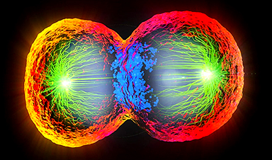
In humans, that means carefully packaging 46 chromosomes and distributing them equally. Before division, each chromosome transforms into a compact, X-shaped structure made of two identical, rod-like copies. But exactly how cells manage to reshape and organize their DNA for this process has remained a mystery.
Now, for the first time, scientists at EMBL have directly visualized this process in high resolution using a new chromatin tracing technique. Their study reveals that during cell division, the long strands of DNA form a series of overlapping loops that push away from one another. This repulsion causes the loops to stack, ultimately giving each chromosome its characteristic rod-like shape.

Looping DNA to Shape Chromosomes
Scientists have long hypothesized the importance of DNA loops in building and maintaining chromosomal structure. First identified in the 1990s, condensins are large protein complexes that bind DNA during cell division and extrude it to create loops of varying sizes. Previous studies from EMBL have shed light on the structural mechanics of this process and their essential role in packing chromosomes into forms that can be easily moved between cells.
In fact, mutations in condensin structure can result in severe chromosome segregation defects and lead to cell death, cancer formation, or rare developmental disorders called 'condensinopathies'.
Solving the DNA Imaging Problem
"However, observing how this looping process occurs on the cellular scale and contributes to chromosome structure is challenging," said Andreas Brunner, postdoc in EMBL Heidelberg's Ellenberg Group and a lead author of the new paper. "This is because methods for visualizing DNA with high resolution are usually chemically harsh and require high temperatures, which together disrupt the native structure of DNA."
Kai Beckwith, a former postdoc in the Ellenberg Group and currently an associate professor at the Norwegian University of Science and Technology (NTNU), set out to solve this problem. Beckwith and colleagues used a method to gently remove one strand of DNA in cells at various stages of cell division, keeping the chromosome structure intact. They could then use targeted sets of DNA-binding labels to observe the nanoscale organization of this uncovered DNA strand. This technique, called LoopTrace, helped the researchers directly observe DNA in dividing cells as it progressively formed loops and folds.
"Andreas and I were now able to visualize the structure of chromosomes as they started to change shape," said Beckwith. "This was crucial for understanding how the DNA was folded by the condensin complexes."
Nested Loops and DNA Compaction
From their data, the scientists realized that during cell division, DNA forms loops in two stages. First, it forms stable large loops, which then subdivide into smaller, short-lived nested loops, increasing the compaction at each stage. Two types of condensin protein complexes enable this process.
To understand how this looping eventually gives rise to rod-shaped chromosomes, the researchers built a computational model based on two simple assumptions. First, as observed, DNA forms overlapping loops – first large and then small – across its length with the help of Condensins. Second, these loops repel each other due to their structure and the chemistry of DNA. When the scientists fed these two assumptions into their model, they found that this was sufficient to give rise to a rod-shaped chromosome structure.
Overlapping Loops Are Key
"We realized that these condensin-driven loops are much larger than previously thought, and that it was very important that the large loops overlap to a significant extent," said Beckwith. "Only these features allowed us to recapitulate the native structure of mitotic chromosomes in our model and understand how they can be segregated during cell division."
In the future, the researchers plan to study this process in more detail, especially to understand how additional factors, such as molecular regulators, affect this compaction process. In 2024, Jan Ellenberg and his team received funding of €3.1 million (~$3.4 million) as an ERC Advanced Grant, to study the folding principles of chromosomes during and following cell division.
A Milestone for Chromosome Biology
"Our newest paper published in the scientific journal Cell marks a milestone in our understanding of how the cell is able to pack chromosomes for their accurate segregation into daughter cells," said Jan Ellenberg, Senior Scientist at EMBL Heidelberg. "It will be the basis to understand the molecular mechanism of rescaling the genome for faithful inheritance and thus rationally predict how errors in this process that underlie human disease could be prevented in the future."
In the meantime, a second study from the Ellenberg Team, led by Andreas Brunner and recently published in the Journal of Cell Biology, shows that the nested loop mechanism is fundamental to the biology of cells, and continues during the cell's growth phase with another family of DNA loop forming protein complexes, called cohesins.
Looping Mechanisms Across Cell Phases
"We were surprised to find that the same core principle of sequential and hierarchical DNA loop formation is used to either tightly pack chromosomes during division into safely movable entities, or to unpack them afterward to read out the information they contain," said Ellenberg. "In the end, small, but key mechanistic differences, such as the non-overlapping nature of cohesin-driven loops compared to the strongly overlapping condensin-driven loops might be sufficient to explain the vast differences that we see in the shape the genome takes in interphase and mitosis under the microscope."
References:
Reference: "Nanoscale DNA tracing reveals the self-organization mechanism of mitotic chromosomes" by Kai Sandvold Beckwith, Andreas Brunner, Natalia Rosalia Morero, Ralf Jungmann and Jan Ellenberg, 24 March 2025, Cell.
DOI: 10.1016/j.cell.2025.02.028
"Quantitative imaging of loop extruders rebuilding interphase genome architecture after mitosis" by Andreas Brunner, Natalia Rosalía Morero, Wanlu Zhang, M. Julius Hossain, Marko Lampe, Hannah Pflaumer, Aliaksandr Halavatyi, Jan-Michael Peters, Kai S. Beckwith and Jan Ellenberg, 9 January 2025, Journal of Cell Biology.
DOI: 10.1083/jcb.202405169
News

Scientists Discover 250+ Genes That Could Lead to New Ways To Prevent Melanoma
The world’s largest study of mole genetics identified hundreds of genes tied to melanoma risk, uncovering potential new drug targets and paving the way for more accurate melanoma screening and prevention. Researchers at QIMR [...]
Breakthrough Diabetes Treatment Reprograms the Immune System
An engineered stem cell therapy reversed new-onset Type 1 diabetes in mice by shifting the immune system away from attacking insulin-producing cells. For more than a century, people with Type 1 diabetes have relied [...]
Taking the world’s temperature: WHO chief spotlights global health emergencies
Taking the world’s temperature on pressing health matters, WHO Director-General Tedros Adhanom Ghebreyesus provided the latest on current global challenges - and successes when it comes to international cooperation. “The outbreaks of hantavirus, Ebola and Marburg all show [...]
Scientists Create Tiny “Mini Livers” That Could One Day Replace Liver Transplants
Engineered tissue grafts could help perform key liver functions and benefit thousands of people living with liver failure. The liver is one of the body’s hardest-working organs, carrying out hundreds of vital jobs, from [...]

NanoMedical Brain/Cloud Interface – Explorations and Implications. A new book from Frank Boehm
New book from Frank Boehm, NanoappsMedical Inc Founder: This book explores the future hypothetical possibility that the cerebral cortex of the human brain might be seamlessly, safely, and securely connected with the Cloud via [...]
Scientists Discover Surprising Way To Help the Brain Recover After Stroke
A new study suggests that strengthening the body’s natural circadian rhythms may help the brain recover after stroke, even when treatment begins days after the injury. Every year, millions of people survive a stroke, [...]

Our books now available worldwide!
Online Sellers other than Amazon, Routledge, and IOPP Indigo Global Health Care Equivalency in the Age of Nanotechnology, Nanomedicine and Artifcial Intelligence Global Health Care Equivalency In The Age Of Nanotechnology, Nanomedicine And Artificial [...]

Younger Generations Are Aging Faster – and It May Be Fueling a Surge in Cancer
Younger generations may be aging biologically faster than those before them, and that shift could help explain rising rates of cancer at younger ages. For decades, cancer was viewed largely as a disease of [...]

Using Cannabis Could Raise Your Stroke Risk by 37%, Massive Study Reveals
Large-scale evidence suggests cannabis, cocaine, and amphetamines may directly raise stroke risk, including in younger adults. As recreational drug use becomes increasingly common, researchers are uncovering evidence that its health consequences may extend far beyond [...]

Could Vitamin C Be the Secret to Keeping Your Brain Younger?
Lower vitamin C levels were linked to reduced brain volume and weaker neural connectivity in older adults, suggesting a potential connection between nutrition and brain health. Could a common vitamin help preserve the brain [...]

This Deadly Disease Was Wiping Out Humans 5,500 Years Ago
A new study suggests plague was already a deadly threat 5,500 years ago, striking small hunter-gatherer communities long before cities and agriculture emerged. For centuries, plague has been remembered as the disease that devastated [...]

China closing in but US leads in biotech quality, commercial reach, survey finds
SAN DIEGO, June 22 (Reuters) - China, which now conducts more clinical drug trials, opens new tab than the U.S., still lags in the quality and commercial reach of its biomedical science, according to a recent survey, opens new [...]

New method generates renewable supply of progenitor immune cells
In a paper published in Cell, a USC Stem Cell-led team reports a new way of generating a renewable and expandable supply of the progenitor cells that give rise to macrophages. These immune cells help [...]

Scientists Just Discovered a Cellular Survival System That Was Never Supposed To Exist
A surprising backup pathway allows cells to make a crucial amino acid when their primary machinery fails. For decades, biologists believed cells had only one way to access a molecule they cannot live without. New [...]

Artificial cells gain porous membranes, enabling lab reactions and drug release
Artificial cells created in the laboratory offer a wide range of potential applications. Until now, however, their membranes—unlike those of real cells—have been virtually impermeable. Researchers at the Max Planck Institute for Polymer Research, [...]

Popular Weight-Loss Drugs Like Ozempic Linked to Lower Breast Cancer Risk
Ozempic and similar weight-loss drugs were linked to a striking 30% reduction in breast cancer risk in a study of more than 110,000 women. Popular weight-loss and diabetes medications such as Ozempic, Wegovy, Mounjaro, [...]