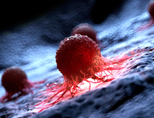
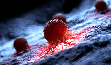
Oregon State University scientists have invented a way to make magnetic nanoparticles that get hotter than any previous nanoparticle, improving their cancer fighting ability.
Findings of the preclinical study led by Oleh Taratula and Olena Taratula were published today in the journal Small Methods.
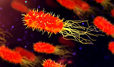
Magnetic nanoparticles have shown anti-cancer potential for years, the scientists said. Once inside a tumor, the particles—tiny pieces of matter as small as one-billionth of a meter—are exposed to an alternating magnetic field. Exposure to the field, a non-invasive process, causes the nanoparticles to heat up, weakening or destroying the cancer cells.
“Magnetic hyperthermia shows great promise for the treatment of many types of cancer,” Olena Taratula said. “Many preclinical and clinical studies have demonstrated its potential to either kill cancer cells directly or enhance their susceptibility to radiation and chemotherapy.”
But at present, magnetic hypothermia can only be used for patients whose tumors are accessible by a hypodermic needle, Oleh Taratula said, and not for people with hard to reach malignancies such as metastatic ovarian cancer.
“With currently available magnetic nanoparticles, the required therapeutic temperatures—above 44 degrees Celsius—can only be achieved by direct injection into the tumor,” he said. “The nanoparticles have only moderate heating efficiency, which means you need a high concentration of them in the tumor to generate enough heat. And numerous studies have shown that only a small percentage of systemically injected nanoparticles accumulate in tumors, making it a challenge to get that high concentration.”
To tackle those problems, the scientists developed a new chemical manufacturing technique that resulted in magnetic nanoparticles with more heating efficiency. They demonstrated in a mouse model that the cobalt-doped nanoparticles will accumulate in metastatic ovarian cancer tumors following low-dose systemic administration, and that when exposed to an alternating magnetic field, the particles can rise in temperature to 50 degrees Celsius.
“To our knowledge, this is the first time it’s been shown that magnetic nanoparticles injected intravenously at a clinically recommended dose are capable of increasing the temperature of cancer tissue above 44 degrees Celsius,” Olena Taratula said. “And we also demonstrated that our novel method could be used for the synthesis of various core-shell nanoparticles. It could serve as a foundation for the development of novel nanoparticles with high heating performance, further advancing systemic magnetic hyperthermia for treating cancer.”
Core-shell nanoparticles have an inner core structure and an outer shell made from different components, she said. Researchers are especially interested in them because of the unique properties that can result from the combination of core and shell material, geometry and design.
In addition to Olena and Oleh Taratula, the collaboration also included College of Pharmacy researchers Youngrong Park, Abraham Moses, Peter Do, Ananiya Demessie, Tetiana Korzun, Fahad Sabei, Conroy Sun, Prem Singh, Fahad Sabei and Hassan Albarqi, as well as Pallavi Dhagat from the Oregon State College of Engineering and researchers from Oregon Health & Science University.
News
Cancer Mystery Solved: Scientists Discover How Melanoma Becomes “Immortal”
Scientists have uncovered a previously overlooked mechanism that may help melanoma cells become effectively “immortal.” Cancer cells face a major problem before they can become deadly: They have to figure out how to stop [...]
How Visual Neurons Organize Thousands of Synaptic Inputs
Summary: A new study uncovered the organizational rules that determine how neurons in the primary visual cortex process information. By imaging both the cell bodies (soma) and the individual synapses (on dendritic spines) of [...]
Scientists Just Found a Surprising Way To Destroy “Forever Chemicals”
Scientists have uncovered a new mechanism that may help break down highly persistent PFAS pollutants. PFAS have earned the nickname “forever chemicals” for a reason. These industrial compounds are so chemically durable that they [...]
Scientists Discover Cheap Material That Kills Deadly Superbugs
A new sulfur-rich antimicrobial polymer shows strong effectiveness against fungal and bacterial pathogens and may offer an affordable solution to antimicrobial resistance. Antimicrobial resistance is creating growing challenges for both healthcare and food production, [...]

What to Know About Cicada, or BA.3.2, the Latest SARS-CoV-2 Variant Under Monitoring
Like periodical cicadas, the insects for which it is nicknamed, SARS-CoV-2 Omicron subvariant BA.3.2 is only just beginning to emerge after lying low for an extended period since it first appeared. Although it was [...]

Scientists Say This Simple Supplement May Actually Reverse Heart Disease
Scientists in Japan say a common supplement may actually help “unclog” certain diseased heart arteries from the inside out. A simple food supplement sold in Japan may have helped reverse a dangerous form of [...]
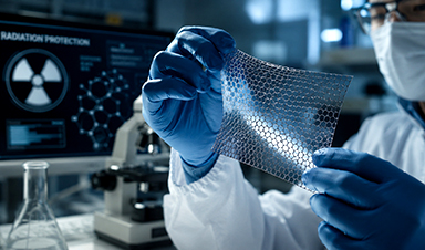
New breakthrough against radiation: Korean Scientists create revolutionary shield with nanotechnology
Korean Scientists develop new nanotechnology material capable of reducing radiation impacts in space missions, hospitals, and power plants. The search for more efficient protection technologies in extreme environments has just gained an important advance. Korean [...]
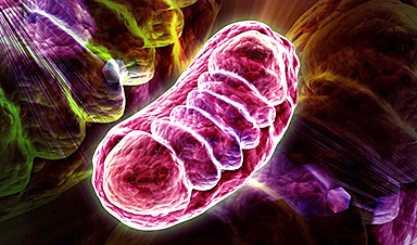
Scientists Just Discovered the Hidden Trick That Keeps Your Cells Alive
A strange bead-like motion inside cells may be the secret to keeping their DNA—and health—in balance. Mitochondria are often described as the power plants of the cell because they produce the energy cells need [...]

Scientists Discover Stem Cells That Could Regrow Teeth and Bone
Scientists just uncovered the cellular “blueprint” that could one day let us regrow real teeth. Researchers at Science Tokyo have uncovered two distinct stem cell lineages that play a central role in forming tooth [...]
Scientists Uncover Fatal Weakness in “Zombie Cells” Linked to Cancer
A newly identified weakness in “zombie” cells may open the door to more precise cancer treatments by turning their own survival strategy against them. A new class of drugs takes advantage of a recently [...]

Bowel and Ovarian Cancers Are Dramatically Rising in Young Adults, Scientists Aren’t Sure Why
Cancer incidence is increasing, especially among younger adults, and current risk factors don’t fully account for the trend. Scientists suggest other underlying causes may be contributing. Cancer patterns in England are shifting in a [...]

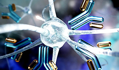
New Immune Pathway Could Supercharge mRNA Cancer Vaccines
A surprising backup system in the immune response to mRNA vaccines may hold the key to more effective cancer treatments. The arrival of mRNA vaccines against SARS-CoV-2 in 2020 marked a turning point in the COVID-19 pandemic. Today, [...]
Scientists Discover “Molecular Switch” That Fuels Alzheimer’s Brain Inflammation
A newly identified trigger of brain inflammation could offer a fresh target for slowing Alzheimer’s progression. The brain has its own built-in immune system that identifies threats and responds to them. In Alzheimer’s disease, growing evidence [...]

Molecular Manufacturing: The Future of Nanomedicine – New book from NanoappsMedical Inc.
This book explores the revolutionary potential of atomically precise manufacturing technologies to transform global healthcare, as well as practically every other sector across society. This forward-thinking volume examines how envisaged Factory@Home systems might enable the cost-effective [...]
Forgotten Medicinal Plant Shows Promise in Fighting Dangerous Superbugs
A traditional medicinal plant, tormentil, shows promise against antibiotic-resistant bacteria in laboratory tests. Its compounds work by limiting bacterial growth and boosting antibiotic performance. Before the development of modern antibiotics, plant-based remedies were commonly [...]

NanoMedical Brain/Cloud Interface – Explorations and Implications. A new book from Frank Boehm
New book from Frank Boehm, NanoappsMedical Inc Founder: This book explores the future hypothetical possibility that the cerebral cortex of the human brain might be seamlessly, safely, and securely connected with the Cloud via [...]