A team of researchers at the Swiss Federal Institute of Technology has developed a high-performance Scanning Ion Conductance Microscope (SICM) using the latest advances in nanopositioning, nanopore fabrication, microelectronics and controls engineering.
Time-resolved scanning allows the 3D visualization of dynamic structures in a eukaryotic cell membrane at nanometer resolutions.
Studying the functions of living cells and organelles at the nanoscale is vital to understanding the causes of disease. Traditional approaches, including electron microscopy, may, unfortunately, damage these cells.
The Swiss researchers developed a SCIM microscope that resolves spatiotemporally diverse three-dimensional processes on a eukaryotic cell membrane at sub-5 nanometer axial resolution. This may offer insights into intracell interactions in the fight against infections and disease.
The Origins of Scanning Probe Microscopy
Studying the intricate functions of living cells at the nanoscale is a unique challenge. Researchers have developed a range of techniques to meet this challenge, including atomic force microscopy (AFM), scanning tunneling microscopy (STM) and Scanning Probe Electrochemistry (SPE).
Scanning probe microscopy (SPM) forms images of surfaces using a probe that scans the specimen. The technique made its first appearance in 1981 in the form of the scanning tunneling microscope, which produces images at atomic resolution by scanning a specimen using a probe.
In scanning probe microscopes, piezoelectric actuators move the probe with atomic-level precision controlled by electronics. The probe raster scans the specimen. It captures discrete data points which are used to form an image. Its manner of scanning is called a mode.
Scanning Ion Conductance Microscopy (SICM) was developed by P.K. Hansma and his colleagues at the University of California in 1989. An electrolyte-containing aqueous medium is a poor conductor.
A SCIM microscope scans a nanoprobe (micro-pipette with a 50 to 100 nm hole) close to the surface of the specimen. As the probe moves across the specimen, ionic currents flow through the pipette. The strengths of these currents vary according to the electrical resistance across the sample’s surface, thus revealing information about its composition.
In the hopping mode described by the Swiss team, however, the nanoprobe is not raster scanned. It moves vertically up and down in a hopping motion.
The probe approaches the specimen at a distance of 25 to 50 nm at specified points and retracts, thus providing discrete points of measurement from which an image is formed. Crucially, the probe never touches the specimen, thus preventing damage to the sample.
SCIM microscopy is, therefore, a powerful tool for capturing the steep changes in a cell’s topography without affecting the sample.
Time-Resolved Scanning Ion Conductance Microscopy
Time-resolved SICM microscopes produce high-resolution profiles of cell shapes and surface characteristics. However, these need to be correlated with biochemical information and changes to the internal organization of the cells.
The Swiss team integrated an inverted optical microscope to a SICM microscope which allowed them to combine recently developed super-resolution microscopy techniques into their approach.
The SICM setup consisted of a custom-built pipette Z-actuator (vertical actuator) integrated into a controlled-atmosphere device, critical for cell viability during imaging.
The imaging of eukaryotic cells requires piezo actuators with a long-range (>10−20 μm). This leads to a trade-off between resonance frequency and the range of the actuator. The team overcame this by adaptively slowing down the pipette’s velocity and applying a gain to the piezo motion as a function of the current interaction curve.
The Z-actuator achieved a wide mechanical displacement amplification of 22 μm scanning range on the cell surface. It was driven by a custom-made piezo controller and integrated with a stepper-motor stage for approaching the sample.
The team used borosilicate and quartz nanopipettes for probing. They were fabricated with a CO2 laser puller with a radius of 20 to 60 nm in size. Quartz capillaries were shrunk to a sub-10-nm radius using electron beam irradiation.
Many cellular processes occur at time scales of minutes or hours and are easily trackable with time-lapse SICM. Subcellular processes, such as endocytosis or infection, however, occur much faster. The Swiss team’s technique combines the capability to address large imaging volumes (up to 220 000 μm3) relatively quickly with high-speed SICM imaging of small details on live cells.
The wide range of measurements possible (Scan sizes from 500 × 500 nm2 to 100 × 100 μm2, imaging speeds from 0.5 s/image to 20 min/image; Number of pixels per image from 1 Kp to 1 Mp; Depth of view of 22 μm with axial resolution below 10 nm) significantly enhances the range of biological studies facilitated by SICM microscopy.
News

China closing in but US leads in biotech quality, commercial reach, survey finds
SAN DIEGO, June 22 (Reuters) - China, which now conducts more clinical drug trials, opens new tab than the U.S., still lags in the quality and commercial reach of its biomedical science, according to a recent survey, opens new [...]

New method generates renewable supply of progenitor immune cells
In a paper published in Cell, a USC Stem Cell-led team reports a new way of generating a renewable and expandable supply of the progenitor cells that give rise to macrophages. These immune cells help [...]

Scientists Just Discovered a Cellular Survival System That Was Never Supposed To Exist
A surprising backup pathway allows cells to make a crucial amino acid when their primary machinery fails. For decades, biologists believed cells had only one way to access a molecule they cannot live without. New [...]

Artificial cells gain porous membranes, enabling lab reactions and drug release
Artificial cells created in the laboratory offer a wide range of potential applications. Until now, however, their membranes—unlike those of real cells—have been virtually impermeable. Researchers at the Max Planck Institute for Polymer Research, [...]

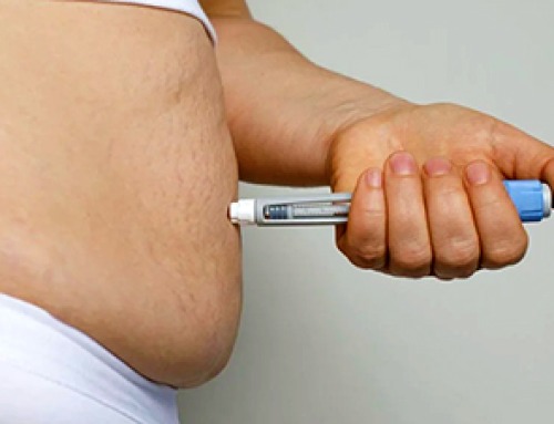
Popular Weight-Loss Drugs Like Ozempic Linked to Lower Breast Cancer Risk
Ozempic and similar weight-loss drugs were linked to a striking 30% reduction in breast cancer risk in a study of more than 110,000 women. Popular weight-loss and diabetes medications such as Ozempic, Wegovy, Mounjaro, [...]

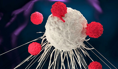
Stanford Scientists Discover Explosive New Type of Immune Cell
Scientists studying the remarkable regenerative abilities of planarian flatworms have uncovered a previously unknown type of immune cell with an unusually destructive defense strategy. What if an immune cell could wipe out nearby threats [...]

Big Pharma-backed SonoThera sounds off with $125M series B for bubble-based genetic delivery
Bay Area biotech SonoThera is bubbling to a clinical boil after raising a $125 million series B with the backing of some of the biggest names in pharma. Vida Ventures led the raise, with the venture [...]

Joint initiative of 5 EU countries calls for ‘unified approach’ to pharma framework amid US drug pricing pressure
With drug pricing pressure building from the U.S., a healthcare-focused consortium of five European countries is calling for a “unified approach” to strengthen Europe’s pharmaceutical framework and access to innovative medicines. Belgium, the Netherlands, [...]

Our books now available worldwide!
Online Sellers other than Amazon, Routledge, and IOPP Indigo Global Health Care Equivalency in the Age of Nanotechnology, Nanomedicine and Artifcial Intelligence Global Health Care Equivalency In The Age Of Nanotechnology, Nanomedicine And Artificial [...]

Molecular Manufacturing: The Future of Nanomedicine – New book from NanoappsMedical Inc.
This book explores the revolutionary potential of atomically precise manufacturing technologies to transform global healthcare, as well as practically every other sector across society. This forward-thinking volume examines how envisaged Factory@Home systems might enable the cost-effective [...]

NanoMedical Brain/Cloud Interface – Explorations and Implications. A new book from Frank Boehm
New book from Frank Boehm, NanoappsMedical Inc Founder: This book explores the future hypothetical possibility that the cerebral cortex of the human brain might be seamlessly, safely, and securely connected with the Cloud via [...]

New book from Nanoappsmedical Inc. – Global Health Care Equivalency
A new book by Frank Boehm, NanoappsMedical Inc. Founder. This groundbreaking volume explores the vision of a Global Health Care Equivalency (GHCE) system powered by artificial intelligence and quantum computing technologies, operating on secure [...]
UCLA Scientists Uncover a “Hidden Weakness” in Some of the World’s Deadliest Cancers
A new study has uncovered an unexpected vulnerability in some of the deadliest cancers. Researchers at UCLA have identified a previously hidden weakness in some of the most aggressive cancers, pointing to a possible new way [...]
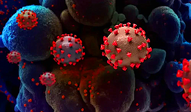
AI-designed universal coronavirus vaccine clears first human trial
Key Takeaways Super-Antigen Technology: Uses AI and machine learning to analyze viral genomes, creating a single vaccine that targets essential features across entire virus families, including coronaviruses and Ebola. Human Trials & Safety: Phase [...]

Researchers Discover a Hidden Vitamin D Problem That Persists Year-Round
A new study suggests that some groups may not experience the expected seasonal boost in vitamin D levels, even during the sunniest months of the year. Many people assume that spending more time outdoors [...]

Researchers Solve the Mystery Behind a Billion-Dollar Dental Implant Disease
Researchers have uncovered why a common and costly dental implant infection often resists antibiotics. Dental implants have helped tens of millions of people regain a full set of stable, functional teeth, something traditional dentures [...]