Cardiovascular disease continues to be the leading cause of death worldwide. But advances in heart-failure therapeutics have stalled, largely due to the difficulty of delivering treatments at the cellular level. Now, a UC Berkeley-led team of researchers may have solved this delivery bottleneck, potentially opening the door to novel, lifesaving treatments.
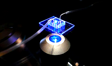
At the core of their new approach is a human cardiac microphysiological system (MPS)—also known as a heart-on-a-chip—that provides a miniaturized model of the human heart, complete with 3D micromuscles. Such devices consist of microfluidic channels, less than the width of a human hair, lined with living human cells. By controlling the fluid flow and other elements, researchers can mimic aspects of the heart’s physiology.
Using their heart-on-a-chip, researchers from UC Berkeley, the Gladstone Institutes and UCSF were able to discover a lipid nanoparticle that could penetrate the dense heart muscle and efficiently deliver its cargo of therapeutic messenger RNA (mRNA) into heart muscle cells, or cardiomyocytes.
Their findings are published in Nature Biomedical Engineering.
Lipid nanoparticles are tiny, spherical particles made of fats that encapsulate therapeutic agents. They are considered the most clinically advanced nonviral transport system for delivering mRNA in gene editing therapies and in vaccines, including the Pfizer-BioNTech and Moderna COVID-19 shots.
However, successfully delivering mRNA to cardiomyocytes hinges on something called endosomal escape, long seen as a challenge in this field. The endosome acts as a cell’s sorting station, and if the therapeutic agent gets stuck there, it will start to degrade. To be effective, the lipid nanoparticle must exit the endosome and enter the cell’s cytoplasm, where it can distribute its mRNA cargo for maximum therapeutic effect.
To tackle this problem, the researchers synthesized lipid nanoparticles with a novel acid-degradable polyethylene glycol coating, with the idea that it could easily diffuse through heart tissue and still leave the endosome. Using their heart-on-a-chip, they then tested different iterations to identify the most effective version for delivering the gene-editing therapy to cardiomyocytes. Later, they tested these same lipid nanoparticles on mouse hearts and recorded similar, positive results.
According to Kevin Healy, co-principal investigator of the study, the researchers’ organ-on-a-chip approach also could allow scientists to more accurately predict test results on live organisms and accelerate advances in mRNA cardiac therapies. The key, he said, is the model’s ability to replicate the complex 3D cellular environments of microtissues better than simple 2D models, which typically consist of a single layer of cells grown in a petri dish.
“Our framework enables faster, animal-sparing identification of effective lipid nanoparticles for safely delivering these therapies,” said Healy, professor of bioengineering and of materials science and engineering at UC Berkeley. “So, by using organ-on-a-chip models to predict heart-targeted delivery and safety, we can potentially accelerate programs for heart failure therapeutics, cardioprotective factors and gene correction, while reducing time and cost to translation.”
More information: Gabriel Neiman et al, A microphysiological system for screening lipid nanoparticle−mRNA complexes predicts in vivo heart transfection efficacy, Nature Biomedical Engineering (2025). DOI: 10.1038/s41551-025-01523-4
Journal information: Nature Biomedical Engineering
News
RNA Recycling Extends Lifespan
Summary: Researchers discovered a biological “trash disposal” mechanism that directly controls how fast we age. While circular RNA has long been known to accumulate in cells as we get older, this study proves for the [...]
Cancer’s Deadly Paradox: How Tumors Break Their Own DNA To Keep Growing
Cancer’s strongest gene switches push DNA into damaging overdrive, creating repeated breaks and repairs that may fuel tumor evolution while exposing possible therapeutic weak spots. A new study indicates that cancer can harm its own genetic [...]

NanoMedical Brain/Cloud Interface – Explorations and Implications. A new book from Frank Boehm
New book from Frank Boehm, NanoappsMedical Inc Founder: This book explores the future hypothetical possibility that the cerebral cortex of the human brain might be seamlessly, safely, and securely connected with the Cloud via [...]

Our books now available worldwide!
Online Sellers other than Amazon, Routledge, and IOPP Indigo Global Health Care Equivalency in the Age of Nanotechnology, Nanomedicine and Artifcial Intelligence Global Health Care Equivalency In The Age Of Nanotechnology, Nanomedicine And Artificial [...]
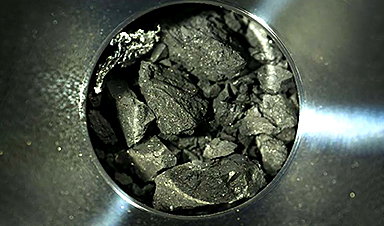
Ryugu asteroid samples contain all DNA and RNA building blocks, bolstering origin-of-life theories
All the essential ingredients to make the DNA and RNA underpinning life on Earth have been discovered in samples collected from the asteroid Ryugu, scientists said Monday. The discovery comes after these building blocks [...]

Is Berberine Really a “Natural Ozempic”?
Often labeled a “natural Ozempic,” berberine is widely discussed as a metabolic aid. Yet research suggests its influence may lie deeper. In recent years, berberine has gained significant attention as a supposed “natural way” [...]
Viagra Ingredient Shows Promise for Rare Childhood Brain Disease in Surprising Study
A rare childhood disease with no approved treatment may have an unexpected new therapeutic candidate. Sildenafil, the active ingredient also sold under the brand name Viagra, may help reduce symptoms in people with Leigh [...]
In a first for China, Neuracle’s implantable brain-computer interface wins approval
In a landmark development, Neuracle Medical Technology has secured the country’s first-ever approval for an implantable brain-computer interface (BCI) system designed to restore hand motor function in patients with spinal cord injuries, in a [...]
A Cambridge Lab Mistake Reveals a Powerful New Way to Modify Drug Molecules
A surprising lab discovery reveals a light-powered way to tweak complex drugs faster, cleaner, and later in development. Researchers at the University of Cambridge have created a new technique for altering complex drug molecules [...]

New book from NanoappsMedical Inc – Molecular Manufacturing: The Future of Nanomedicine
This book explores the revolutionary potential of atomically precise manufacturing technologies to transform global healthcare, as well as practically every other sector across society. This forward-thinking volume examines how envisaged Factory@Home systems might enable the cost-effective [...]
Scientists Discover Simple Saliva Test That Reveals Hidden Diabetes Risk
Researchers have identified a potential new way to assess metabolic health using saliva instead of blood. High insulin levels in the blood, known as hyperinsulinemia, can reveal metabolic problems long before obvious symptoms appear. It is [...]

One Nasal Spray Could Protect Against COVID, Flu, Pneumonia, and More
A single nasal spray vaccine may one day protect against viruses, pneumonia, and even allergies. For decades, scientists have dreamed of creating a universal vaccine capable of protecting against many different pathogens. The idea [...]
New AI Model Predicts Cancer Spread With Incredible Accuracy
Scientists have developed an AI system that analyzes complex gene-expression signatures to estimate the likelihood that a tumor will spread. Why do some tumors spread throughout the body while others remain confined to their [...]
Scientists Discover DNA “Flips” That Supercharge Evolution
In Lake Malawi, hundreds of species of cichlid fish have evolved with astonishing speed, offering scientists a rare opportunity to study how biodiversity arises. Researchers have identified segments of “flipped” DNA that may allow fish to adapt rapidly [...]
Scientists Discover Why Some COVID Survivors Still Can’t Taste Food Years Later
A new study provides the first direct biological evidence explaining why some people continue to experience taste loss long after recovering from COVID-19. Researchers have uncovered specific biological changes in taste buds that could help [...]

Catching COVID significantly raises the risk of developing kidney disease, researchers find
Catching Covid significantly raises the risk of developing deadly kidney disease, research has shown. The virus was found to increase the chances that patients will develop the incurable condition by around 50 per cent. [...]