A deadly, drug-resistant hospital fungus may finally have a weakness—and scientists think they’ve found it.
Researchers have identified a genetic process that could open the door to new treatments for a dangerous fungal infection that has repeatedly forced hospital intensive care units to close. The discovery offers fresh hope against a pathogen that has been difficult to control and even harder to treat.
Candida auris poses a serious threat to people who are already critically ill, making hospitals particularly vulnerable to outbreaks. Although the fungus can exist harmlessly on the skin of many people, patients who rely on ventilators face a much higher risk of infection. Once it takes hold, the disease kills around 45 per cent of those infected and can withstand every major class of antifungal medication. This combination has made it extremely challenging to eliminate from hospital wards once it spreads.
A global health threat on the rise
Candida auris was first identified in 2008, and scientists still do not know where it originally came from. Since its discovery, outbreaks have been reported in more than 40 countries, including the UK. The fungus, also known as Candidozyma auris, is now recognized as a global health threat and appears on the World Health Organization’s critical priority fungal pathogens list. In the UK, case numbers have continued to climb over time.
Studying infection in a living host
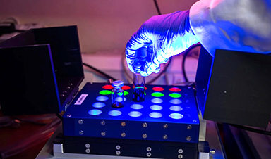
For the first time, a team at the University of Exeter has closely examined how Candida auris activates its genes during infection using an innovative experimental model based on fish larvae. The research was published in the Nature portfolio journal Communications Biology and received support from Wellcome, the Medical Research Council (MRC), and the National Center for Replacement, Reduction and Refinement (NC3Rs).
The results suggest a promising path toward identifying a biological target that could be used to develop new antifungal drugs or adapt existing ones, provided the same genetic activity occurs during infection in humans.
The work was co-led by NIHR Clinical Lecturer Hugh Gifford from the University of Exeter’s MRC Center for Medical Mycology (CMM). He said: “Since it emerged, Candida auris has wreaked havoc where it takes hold in hospital intensive care units. It can be deadly for vulnerable patients, and health trusts have spent millions on the difficult job of eradication. We think our research may have revealed an Achilles heel in this lethal pathogen during active infection, and we urgently need more research to explore whether we can find drugs that target and exploit this weakness.”
Why a new model was needed
One long-standing challenge in studying Candida auris is its ability to survive high temperatures. Combined with its unusually strong tolerance to salt, this has led some scientists to speculate that it may have originated in tropical oceans or marine animals. These traits have also made it harder to study using traditional laboratory models.
To overcome this, the Exeter researchers developed a new system using Arabian killifish, whose eggs are able to survive at human body temperature. This allowed the team to observe the infection process in a living host under realistic conditions.
Genetic clues to survival and spread
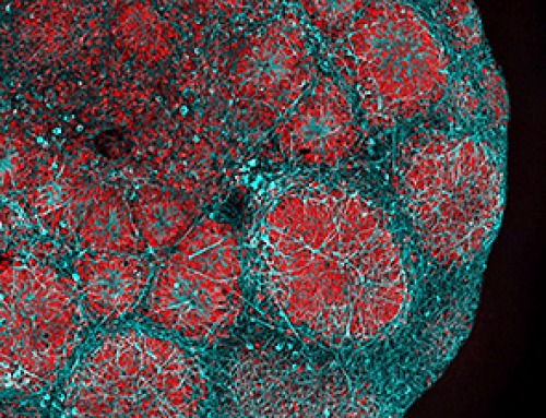
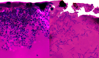
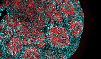
During the study, the researchers observed that Candida auris can shift into elongated fungal structures called filaments, which may help it search for nutrients inside the host.
They also tracked which genes were turned on and off during infection, highlighting potential weaknesses. Several of the activated genes are involved in producing nutrient pumps that capture iron-scavenging molecules and pull iron into fungal cells. Iron is essential for survival, and this dependence could represent a critical vulnerability.
Co-senior author Dr. Rhys Farrer from the University of Exeter’s MRC Centre for Medical Mycology said: “Until now, we’ve had no idea what genes are active during infection of a living host. We now need to find out if this also occurs during human infection. The fact that we found genes are activated to scavenge iron gives clues to where Candida auris may originate, such as an iron-poor environment in the sea. It also gives us a potential target for new and already existing drugs.”
Hope for future treatments
Dr. Gifford, who also works as a resident physician in intensive care and respiratory medicine at the Royal Devon & Exeter Hospital, emphasized the potential medical impact of the findings. He said: “While there are a number of research steps to go through yet, our finding could be an exciting prospect for future treatment. We have drugs that target iron scavenging activities. We now need to explore whether they could be repurposed to stop Candida auris from killing humans and closing down hospital intensive care units.”
The Arabian killifish larvae model was developed with support from an NC3Rs project grant as an alternative to using mouse and zebrafish models, which are commonly employed to study how pathogens interact with their hosts.
Dr. Katie Bates, NC3Rs Head of Research Funding, said: “This new publication demonstrates the utility of the replacement model to study Candida auris infection and enable unprecedented insights into cellular and molecular events in live infected hosts. This is a brilliant example of how innovative alternative approaches can overcome key limitations of traditional animal studies.”
Reference: “Xenosiderophore transporter gene expression and clade-specific filamentation in Candida auris killifish (Aphanius dispar) infection” 19 December 2025, Communications Biology.
DOI: 10.1038/s42003-025-09321-z
News

NanoMedical Brain/Cloud Interface – Explorations and Implications. A new book from Frank Boehm
New book from Frank Boehm, NanoappsMedical Inc Founder: This book explores the future hypothetical possibility that the cerebral cortex of the human brain might be seamlessly, safely, and securely connected with the Cloud via [...]

Our books now available worldwide!
Online Sellers other than Amazon, Routledge, and IOPP Indigo Global Health Care Equivalency in the Age of Nanotechnology, Nanomedicine and Artifcial Intelligence Global Health Care Equivalency In The Age Of Nanotechnology, Nanomedicine And Artificial [...]

Ryugu asteroid samples contain all DNA and RNA building blocks, bolstering origin-of-life theories
All the essential ingredients to make the DNA and RNA underpinning life on Earth have been discovered in samples collected from the asteroid Ryugu, scientists said Monday. The discovery comes after these building blocks [...]

Is Berberine Really a “Natural Ozempic”?
Often labeled a “natural Ozempic,” berberine is widely discussed as a metabolic aid. Yet research suggests its influence may lie deeper. In recent years, berberine has gained significant attention as a supposed “natural way” [...]
Viagra Ingredient Shows Promise for Rare Childhood Brain Disease in Surprising Study
A rare childhood disease with no approved treatment may have an unexpected new therapeutic candidate. Sildenafil, the active ingredient also sold under the brand name Viagra, may help reduce symptoms in people with Leigh [...]

In a first for China, Neuracle’s implantable brain-computer interface wins approval
In a landmark development, Neuracle Medical Technology has secured the country’s first-ever approval for an implantable brain-computer interface (BCI) system designed to restore hand motor function in patients with spinal cord injuries, in a [...]
A Cambridge Lab Mistake Reveals a Powerful New Way to Modify Drug Molecules
A surprising lab discovery reveals a light-powered way to tweak complex drugs faster, cleaner, and later in development. Researchers at the University of Cambridge have created a new technique for altering complex drug molecules [...]

New book from NanoappsMedical Inc – Molecular Manufacturing: The Future of Nanomedicine
This book explores the revolutionary potential of atomically precise manufacturing technologies to transform global healthcare, as well as practically every other sector across society. This forward-thinking volume examines how envisaged Factory@Home systems might enable the cost-effective [...]

Scientists Discover Simple Saliva Test That Reveals Hidden Diabetes Risk
Researchers have identified a potential new way to assess metabolic health using saliva instead of blood. High insulin levels in the blood, known as hyperinsulinemia, can reveal metabolic problems long before obvious symptoms appear. It is [...]

One Nasal Spray Could Protect Against COVID, Flu, Pneumonia, and More
A single nasal spray vaccine may one day protect against viruses, pneumonia, and even allergies. For decades, scientists have dreamed of creating a universal vaccine capable of protecting against many different pathogens. The idea [...]
New AI Model Predicts Cancer Spread With Incredible Accuracy
Scientists have developed an AI system that analyzes complex gene-expression signatures to estimate the likelihood that a tumor will spread. Why do some tumors spread throughout the body while others remain confined to their [...]

Scientists Discover DNA “Flips” That Supercharge Evolution
In Lake Malawi, hundreds of species of cichlid fish have evolved with astonishing speed, offering scientists a rare opportunity to study how biodiversity arises. Researchers have identified segments of “flipped” DNA that may allow fish to adapt rapidly [...]

Scientists Discover Why Some COVID Survivors Still Can’t Taste Food Years Later
A new study provides the first direct biological evidence explaining why some people continue to experience taste loss long after recovering from COVID-19. Researchers have uncovered specific biological changes in taste buds that could help [...]

Catching COVID significantly raises the risk of developing kidney disease, researchers find
Catching Covid significantly raises the risk of developing deadly kidney disease, research has shown. The virus was found to increase the chances that patients will develop the incurable condition by around 50 per cent. [...]

New Toothpaste Stops Gum Disease Without Harming Healthy Bacteria
Researchers have developed a targeted approach to combat periodontitis without disrupting the natural balance of the oral microbiome. The innovation could reshape how gum disease is treated while preserving beneficial bacteria. The human mouth [...]

Plastic Without End: Are We Polluting the Planet for Eternity?
The Kunming Montreal Global Biodiversity Framework calls for the elimination of plastic pollution by 2030. If that goal has been clearly set, why have meaningful measures that create real change still not been implemented? [...]