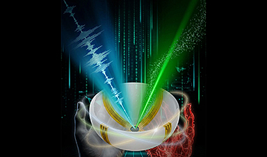
A newly developed imaging method blends ultrasound and photoacoustics to capture both tissue structure and blood-vessel function in 3D.
By blending two powerful imaging methods, researchers from Caltech and USC have developed a new way to see inside the human body with unprecedented speed and detail. The technique produces three-dimensional, full-color images that show not only the shape of soft tissues but also how blood vessels are functioning in real time. In early demonstrations, the researchers successfully imaged several different parts of the human body, highlighting the versatility of the approach.
This combined imaging method could significantly improve how doctors detect and study disease. Potential applications include more precise breast tumor imaging, new ways to track nerve damage caused by diabetes, and advanced tools for observing brain structure alongside blood flow. The work suggests a path toward medical scans that are both more informative and easier to repeat over time.
The researchers describe the new technology in a paper published in Nature Biomedical Engineering.
Medical imaging often requires tradeoffs between speed, cost, and the type of information that can be captured. Ultrasound, one of the most widely used techniques, is fast, inexpensive, and well suited for visualizing the structure of tissues. However, it typically provides only two-dimensional views and cannot easily capture a wide area or reveal detailed information about blood chemistry or flow.
Photoacoustic imaging addresses some of those gaps but introduces others. In this approach, laser light is sent into the body, where it is absorbed by molecules in blood vessels. That absorption generates sound waves that can be detected and translated into images. Because different molecules absorb light in distinct ways, photoacoustic imaging can display blood vessels in optical color—allowing for visualization of how blood moves through arteries and veins. On its own, however, the technique does not provide enough structural detail to fully map surrounding tissues.
Other advanced imaging tools, such as computed tomography (CT) scanning and magnetic resonance imaging (MRI), can deliver detailed views of anatomy, but they come with notable downsides. These methods can be costly, may require contrast agents, sometimes involve exposure to ionizing radiation, or take too long to be practical for frequent monitoring or bedside use.
Combining Ultrasound and Photoacoustics
To overcome these limitations, the researchers developed RUS-PAT (rotational ultrasound tomography, RUST, combined with photoacoustic tomography, PAT). PAT was first developed more than two decades ago by Lihong Wang, the Bren Professor of Medical Engineering and Electrical Engineering and the Andrew and Peggy Cherng Medical Engineering Leadership Chair at Caltech.
In PAT, molecules that absorb light respond to short laser pulses by vibrating, which generates acoustic signals. These signals can then be detected and processed to form detailed, high-resolution images.
Wang, who is also the executive officer for medical engineering at Caltech, says his group's aim with the current work was to combine the benefits of PAT with ultrasound. "But it's not like one plus one," he says. "We needed to find an optimal way of combining the two technologies."
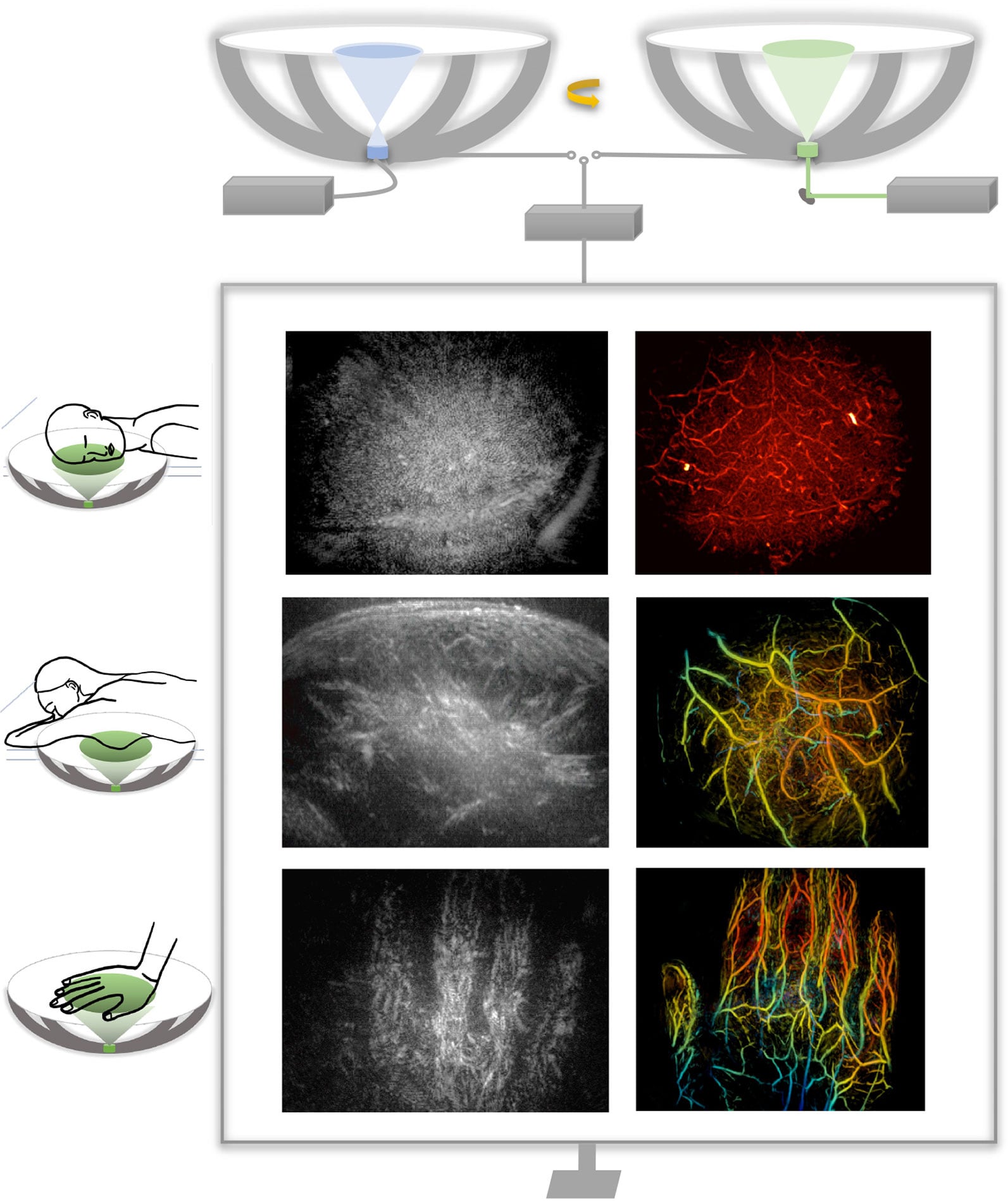
Ultrasound typically uses many transducers to both generate and receive ultrasound waves, and combining this process directly with PAT would be too complex and expensive for widespread use. PAT, meanwhile, only requires the detection of ultrasound, and that gave Wang an idea. "I thought, 'Wait, can we just mimic light excitation of ultrasound waves in photoacoustic tomography, but do it ultrasonically?'" PAT allows laser light to diffuse within the tissue, ultimately triggering the production of measurable ultrasound waves. Similarly, Wang figured, they could use a single wide-field ultrasound transducer to broadcast an ultrasonic wave broadly into the tissue.
They could then use the same detectors to measure the resulting waves for both modalities. In the new system, a small number of arc-shaped detectors are rotated around a central point, allowing it to behave like a full hemispheric detector but at a fraction of the complexity and cost.
Demonstrated Clinical Potential
"The novel combination of acoustic and photoacoustic techniques addresses many of the key limitations of widely used medical-imaging techniques in current clinical practice, and, importantly, the feasibility for human application has been demonstrated here in multiple contexts," says Dr. Charles Y. Liu, an author of the paper who is a visiting associate in biology and biological engineering at Caltech. Liu is also a professor at the Keck School of Medicine of USC, director of USC's Neurorestoration Center, and chair of neurosurgery at the Rancho Los Amigos National Rehabilitation Center.
The RUS-PAT technique could potentially be used in any region of the body to which light can be delivered, and for applications where clinicians or researchers would benefit from the synergistic imaging of both the morphology and color-related function. For example, RUS-PAT could improve breast-tumor imaging, giving physicians the ability to know a tumor's exact location and surroundings as well as its pathology and physiology. It could also help doctors monitor the nerve damage caused by diabetic neuropathy by providing an all-in-one way to monitor oxygen supply along with morphology. Wang says the technique could also be useful in brain imaging, allowing scientists to observe the structural details of the brain while also being able to observe hemodynamics.
Currently, the system can scan to a depth of about 4 centimeters. Light can also be delivered endoscopically, potentially making deeper tissues accessible to the new technology. A RUS-PAT scan can be performed in less than one minute.
The current setup involves a scanning system with ultrasound transducers and laser housed underneath a bed. It has been demonstrated on human volunteers and patients and is in the early stages of translational development.
Reference: "Rotational ultrasound and photoacoustic tomography of the human body" by Yang Zhang, Shuai Na, Jonathan J. Russin, Karteekeya Sastry, Li Lin, Junfu Zheng, Yilin Luo, Xin Tong, Yujin An, Peng Hu, Konstantin Maslov, Tze-Woei Tan, Charles Y. Liu and Lihong V. Wang, 16 January 2026, Nature Biomedical Engineering.
DOI: 10.1038/s41551-025-01603-5
The work was supported by funding from the National Institutes of Health.
News
Scientists Discover the Brain Protein That Helps Alzheimer’s Spread Through the Brain
Scientists have identified a brain protein that may help Alzheimer’s spread, revealing a potential new target for slowing the disease’s progression. Alzheimer’s disease is closely linked to the accumulation of a toxic form of the protein [...]
How Immune Dysregulation Contributes to Psychiatric Disorders
Introduction Growing evidence suggests that disruptions in immune function may play an important role in the development and progression of psychiatric disorders. However, immune mechanisms probably contribute more strongly in some patients than others, [...]
Electrostatic Discharge Boosts Triboelectric Nanogenerator Current and Enables DC Output
Controlled electrical discharges could enable triboelectric nanogenerators to achieve higher peak currents, extending nano-enabled energy harvesting into chemical processing and self-powered sensing. Paper: Electrostatic discharge as a breakthrough strategy for triboelectric nanogenerators. A new review [...]
Swiss laboratory uses old drugs against rare diseases
Researchers at the University of Geneva are combing through collections of approved drugs to find new therapies for rare diseases – with some success. This approach is gaining traction around the world, while pharmaceutical [...]
Nanozyme Aptasensors Show Promise for Faster Food, Health, and Environmental Testing
By pairing robust artificial enzymes with highly selective aptamers, nanozyme aptasensors could help detect disease biomarkers, pathogens, and contaminants faster, but the review shows that real-world deployment still depends on overcoming matrix interference, biofouling, [...]

Paralyzed Man Feels Sensation Again With Brain Stimulation Device
Aneuroprosthetic system has allowed a man with paralysis to grasp and lift objects and feel touch again. The device helped 42-year-old Keith Thomas of Massapequa, New York, who was paralyzed from the chest down [...]
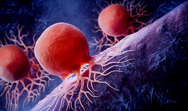
Global Cancer Cases Could Surge 67% by 2050, New Report Warns
New data reveal major geographic disparities and highlight the urgent need for global action on prevention, early detection, and equitable access to treatment. For roughly one in five people worldwide, cancer will become part [...]
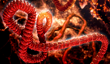
A Deadly Ebola-Like Virus Is Spreading. Are We Ready?
BU virologist Nancy Sullivan says the Bundibugyo outbreak in the Democratic Republic of the Congo underscores the need for broader outbreak preparedness. The death of a nurse marked the moment health officials recognized that [...]
Why Most Animal Viruses Never Become Human Pandemics
From receptor mismatch to risky human-animal interfaces, this article explains why spillover is common but true pandemic emergence remains rare. Introduction Humans are constantly exposed to animal viruses through farming, wildlife contact, and the [...]

Stem cell organoids repair heart microvessels in coronary artery disease models
A Stanford University team has shown that vascular organoids derived from human stem cells can repair the heart’s microvessel network in pigs with ischaemic heart disease – a proof-of-concept advancement that could open new therapeutic [...]

Goodbye GP waiting rooms, hello prevention at home
Prevention is suddenly everywhere in NHS reform. The recent £340m community pharmacy deal is moving more services onto the high street. Community Diagnostic Centres are being expanded, and the Neighbourhood Health Framework sets out [...]
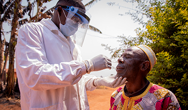
Ebola control is weakened by mistrust and cultural insensitivity
Effective response depends on cooperation with communities and frontline workers, writes Zaeem ul Haq The current Bundibugyo Ebola outbreak in the Democratic Republic of the Congo (DRC) and Uganda is exposing dangerous gaps in [...]
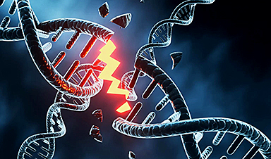
Building the Brain Requires Millions of Dangerous DNA Breaks
Scientists discovered that building a healthy brain involves an unexpected step: young neurons routinely break and rapidly repair their own DNA. As the brain develops, newly formed nerve cells must travel through tightly packed tissue [...]

One Tiny Change May Explain How Viruses Jump From Bats to Humans
Scientists found that one tiny genetic change may determine whether a bat virus stays in bats or becomes a human threat. Most infectious disease outbreaks begin when a virus or other pathogen crosses from animals into [...]
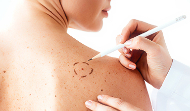
Scientists Discover 250+ Genes That Could Lead to New Ways To Prevent Melanoma
The world’s largest study of mole genetics identified hundreds of genes tied to melanoma risk, uncovering potential new drug targets and paving the way for more accurate melanoma screening and prevention. Researchers at QIMR [...]
Breakthrough Diabetes Treatment Reprograms the Immune System
An engineered stem cell therapy reversed new-onset Type 1 diabetes in mice by shifting the immune system away from attacking insulin-producing cells. For more than a century, people with Type 1 diabetes have relied [...]