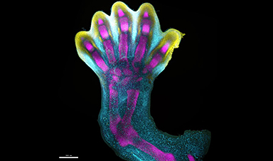
Human fingers and toes don't grow outward as you might expect. Instead, our dexterous digits are 'sculpted' within a larger foundational bud.
Now the first human cell atlas of early limb development has at last revealed in exquisite detail exactly how that happens.
Prior to this, our understanding of vertebrate limb development has been largely based on model organisms, such as mice and chicken embryos, and lab-grown stem cells.
Although humans share some similarities with other vertebrates, their biology obviously diverges from ours.
The details of early limb formation have also been rendered a little fuzzy by technological limitations, now surpassed, and restrictions on the use of human embryos for research beyond 14 days, a rule that has been relaxed under strict ethical provisions.
The picture constructed so far had limbs initially emerging as shapeless limb buds protruding from the sides of the embryonic body. Eight weeks later, if all goes to plan, those pouches have transformed into anatomically distinct, recognizable limbs, complete with fingers and toes.
It's a remarkable process in early embryonic development that produces arguably one of our most defining human features: our long, slender, opposable thumbs.
In 2014, scientists described how specific molecules expressed at precise moments in embryonic development moulded the formation of fingers and toes, although those predictions were based on simulations of experimental data.
Now, an international team led by cell biologist Bao Zhang at Sun Yat-sen University in China, has colored in that process in exquisite detail, by analyzing thousands of single cells from donated embryonic tissues that were between 5 and 9 weeks of development.
")
![]()
"We identified 67 distinct cell clusters from 125,955 captured single cells, and spatially mapped them across four first trimester timepoints to shed new light on limb development," the team writes in their published paper.
"In doing so, we uncovered several new cell states," they add.
"What we reveal is a highly complex and precisely regulated process," says Hongbo Zhang, senior author and cell biologist from Sun Yat-sen University in China.
"It is like watching a sculptor at work, chiseling away at a block of marble to reveal a masterpiece. In this case, nature is the sculptor, and the result is the incredible complexity of our fingers and toes."
As you can see in the video below, the researchers mapped gene expression patterns to see how those genetic instructions shaped how digits formed.
From hazy beginnings, the expression of IRX1 (represented in aqua in the video below), a gene critical for digit formation, and SOX9 (represented in magenta in the video), a gene essential for skeletal development, overlap in five distinct lengths within the developing limb.
At around 7 weeks of development, programmed cell death instructions are switched on in the undifferentiated cells congregating between these lengths (associated with the expression of MSX1, represented in yellow in the video), and well-defined fingers and toes are revealed.
Like a block of marble being sculpted into a masterpiece by the expression of these genes, our fingers and toes are chiseled out from tip to base as unneeded cells recede.
Small irregularities in this process can lead to limb deformities, which 1 in 500 people are born with – making them some of the most frequently reported syndromes at birth.
The researchers also mapped the expression of genes linked with congenital conditions, such as short fingers (brachydactyly) or webbed digits (syndactyly), to get a better sense of where limb development gets off course.
"For the first time, we have been able to capture the remarkable process of limb development down to single-cell resolution in space and time," says Sarah Teichmann, senior author and computational biologist at the Wellcome Sanger Institute.
She says creating single-cell atlases is "deepening our understanding of how anatomically complex structures form, helping us uncover the genetic and cellular processes behind healthy human development, with many implications for research and healthcare."
Importantly, the researchers also showed that limb formation in humans and mice does follow similar trajectories, with some differences in activated genes and cell types.
The study has been published in Nature.
News

GHCE Concept
From the preface of the book Global Health Care Equivalency in the Age of Nanotechnology, Nanomedicine and Artificial Intelligence, Edited by Frank Boehm: Since the publication of my first book (Nanomedical Device and Systems [...]

Healthcare Headlines: Challenges and Advances in 2026
Health-related updates reveal financial adjustments by Universal Health Services due to Medicaid reimbursement uncertainties, significant pollution-linked health concerns from French-British oil firm Perenco in Congo, drug trial setbacks, potential restructuring at major medical firms, [...]
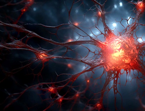
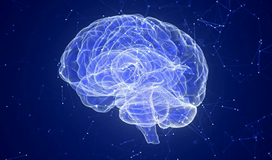
Scientists Discover the Brain Protein That Helps Alzheimer’s Spread Through the Brain
Scientists have identified a brain protein that may help Alzheimer’s spread, revealing a potential new target for slowing the disease’s progression. Alzheimer’s disease is closely linked to the accumulation of a toxic form of the protein [...]
How Immune Dysregulation Contributes to Psychiatric Disorders
Introduction Growing evidence suggests that disruptions in immune function may play an important role in the development and progression of psychiatric disorders. However, immune mechanisms probably contribute more strongly in some patients than others, [...]
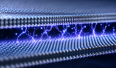
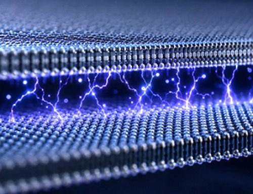
Electrostatic Discharge Boosts Triboelectric Nanogenerator Current and Enables DC Output
Controlled electrical discharges could enable triboelectric nanogenerators to achieve higher peak currents, extending nano-enabled energy harvesting into chemical processing and self-powered sensing. Paper: Electrostatic discharge as a breakthrough strategy for triboelectric nanogenerators. A new review [...]
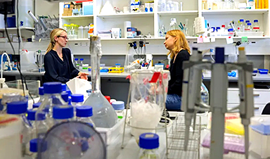
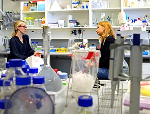
Swiss laboratory uses old drugs against rare diseases
Researchers at the University of Geneva are combing through collections of approved drugs to find new therapies for rare diseases – with some success. This approach is gaining traction around the world, while pharmaceutical [...]
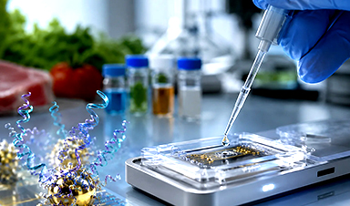
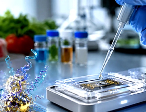
Nanozyme Aptasensors Show Promise for Faster Food, Health, and Environmental Testing
By pairing robust artificial enzymes with highly selective aptamers, nanozyme aptasensors could help detect disease biomarkers, pathogens, and contaminants faster, but the review shows that real-world deployment still depends on overcoming matrix interference, biofouling, [...]

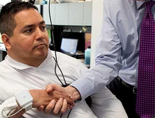
Paralyzed Man Feels Sensation Again With Brain Stimulation Device
Aneuroprosthetic system has allowed a man with paralysis to grasp and lift objects and feel touch again. The device helped 42-year-old Keith Thomas of Massapequa, New York, who was paralyzed from the chest down [...]
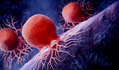
Global Cancer Cases Could Surge 67% by 2050, New Report Warns
New data reveal major geographic disparities and highlight the urgent need for global action on prevention, early detection, and equitable access to treatment. For roughly one in five people worldwide, cancer will become part [...]
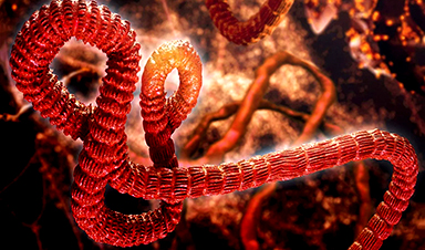
A Deadly Ebola-Like Virus Is Spreading. Are We Ready?
BU virologist Nancy Sullivan says the Bundibugyo outbreak in the Democratic Republic of the Congo underscores the need for broader outbreak preparedness. The death of a nurse marked the moment health officials recognized that [...]
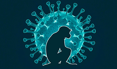
Why Most Animal Viruses Never Become Human Pandemics
From receptor mismatch to risky human-animal interfaces, this article explains why spillover is common but true pandemic emergence remains rare. Introduction Humans are constantly exposed to animal viruses through farming, wildlife contact, and the [...]
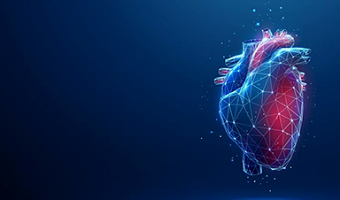
Stem cell organoids repair heart microvessels in coronary artery disease models
A Stanford University team has shown that vascular organoids derived from human stem cells can repair the heart’s microvessel network in pigs with ischaemic heart disease – a proof-of-concept advancement that could open new therapeutic [...]

Goodbye GP waiting rooms, hello prevention at home
Prevention is suddenly everywhere in NHS reform. The recent £340m community pharmacy deal is moving more services onto the high street. Community Diagnostic Centres are being expanded, and the Neighbourhood Health Framework sets out [...]
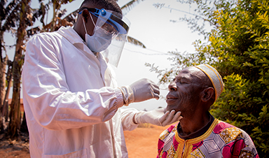
Ebola control is weakened by mistrust and cultural insensitivity
Effective response depends on cooperation with communities and frontline workers, writes Zaeem ul Haq The current Bundibugyo Ebola outbreak in the Democratic Republic of the Congo (DRC) and Uganda is exposing dangerous gaps in [...]
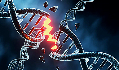
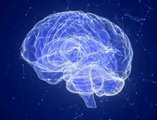
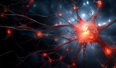
Building the Brain Requires Millions of Dangerous DNA Breaks
Scientists discovered that building a healthy brain involves an unexpected step: young neurons routinely break and rapidly repair their own DNA. As the brain develops, newly formed nerve cells must travel through tightly packed tissue [...]

One Tiny Change May Explain How Viruses Jump From Bats to Humans
Scientists found that one tiny genetic change may determine whether a bat virus stays in bats or becomes a human threat. Most infectious disease outbreaks begin when a virus or other pathogen crosses from animals into [...]