A study in Scientific Reports evaluated a photoacoustic polydopamine-indocyanine green (PDA-ICG) nanoprobe for detecting senescent cells. Senescent cells play a role in tumor progression and therapeutic resistance, with potential adverse effects such as inflammation and tissue disruption. The PDA-ICG nanoprobe offers a method for identifying these cells, with implications for cancer diagnostics and treatment.
Background
Cellular senescence is a stable cell cycle arrest triggered by stressors such as DNA damage, oxidative stress, and oncogenic signaling. Senescent cells produce pro-inflammatory cytokines, growth factors, and proteases, collectively known as the senescence-associated secretory phenotype (SASP).
This phenomenon contributes to tumor progression and age-related diseases. Accurately identifying and visualizing these cells in vivo is crucial for understanding their role in cancer biology and developing targeted therapies.
Traditional methods for detecting senescent cells, such as β-galactosidase staining and immunohistochemistry, have limitations in terms of specificity and sensitivity. The introduction of advanced imaging techniques, particularly those utilizing nanoprobes, offers a promising avenue for enhancing the detection of senescent cells.
The PDA-ICG nanoprobe combines the photothermal properties of polydopamine with the fluorescence of indocyanine green, enabling both photoacoustic imaging and fluorescence imaging. This dual functionality is expected to improve the visualization of senescent cells.
The Current Study
The study used experimental techniques to evaluate the performance of the PDA-ICG nanoprobe. Human cancer cell lines, A549 and SK-MEL-103, were cultured and treated with varying concentrations of PDA-ICG to assess cell viability and nanoprobe internalization.
The MTS assay measured cell viability after treatment, and flow cytometry assessed nanoprobe internalization in live cells. After treatment, cells were washed to remove excess probes and stained with DAPI for flow cytometric analysis. Data were processed using FlowJo software to identify live, single-cell populations with internalized nanoprobe.
Cells were treated with chemotherapeutic agents, cisplatin and palbociclib, for a specified duration. After drug removal, the cells were stained with β-galactosidase to identify senescent cells. RNA extraction and quantitative real-time PCR (RT-qPCR) were performed to measure the expression levels of senescence-associated genes.
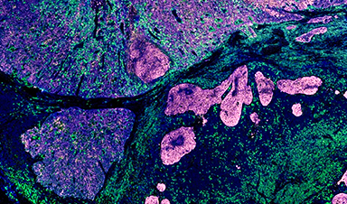
Western blotting was conducted to analyze protein expression related to senescence, including p21 and pRb. Confocal microscopy was utilized to visualize the cellular localization of the PDA-ICG nanoprobe and assess its potential for imaging senescent cells.
Results and Discussion
The results showed that the PDA-ICG nanoprobe was successfully internalized into cancer cells, with flow cytometry confirming significantly higher uptake in treated cells than controls. The MTS assay indicated no adverse effects on cell viability at the tested concentrations, supporting its potential for safe in vivo application.
The study also found that treatment with cisplatin and palbociclib successfully induced senescence in the respective cell lines, as evidenced by increased β-galactosidase activity. The expression of senescence-associated genes was significantly elevated in treated cells, further confirming the induction of senescence.
Confocal microscopy highlighted the PDA-ICG nanoprobe’s imaging capabilities, revealing distinct localization patterns within the cells. The nanoprobe’s dual imaging modality allowed for more precise visualization of senescent cells than traditional methods. The findings suggest that the PDA-ICG nanoprobe could serve as a valuable tool for studying the dynamics of senescence in cancer and other diseases.
The ability to visualize senescent cells in real time may facilitate the development of targeted therapies that eliminate these cells from the tumor microenvironment, potentially improving patient outcomes.
Conclusion
The study successfully demonstrated the utility of the PDA-ICG nanoprobe for detecting senescent cells in cancer. Combining the advantages of photoacoustic and fluorescence imaging, this innovative approach offers a promising strategy for enhancing the visualization of senescence in vivo. The findings underscore the importance of accurately identifying senescent cells in the context of cancer biology and therapeutic interventions.
Future research should focus on optimizing the nanoprobe for clinical applications and exploring its potential in various cancer models. The ability to monitor senescence dynamically could lead to significant advancements in cancer diagnostics and treatment, ultimately contributing to improved patient care and outcomes.
Journal Reference
Hartono, M., et al. (2024). Photoacoustic polydopamine-indocyanine green (PDA-ICG) nanoprobe for detection of senescent cells. Scientific Reports. DOI: 10.1038/s41598-024-79667-7, https://www.nature.com/articles/s41598-024-79667-7

News

Our books now available worldwide!
Online Sellers other than Amazon, Routledge, and IOPP Indigo Global Health Care Equivalency in the Age of Nanotechnology, Nanomedicine and Artifcial Intelligence Global Health Care Equivalency In The Age Of Nanotechnology, Nanomedicine And Artificial [...]

Younger Generations Are Aging Faster – and It May Be Fueling a Surge in Cancer
Younger generations may be aging biologically faster than those before them, and that shift could help explain rising rates of cancer at younger ages. For decades, cancer was viewed largely as a disease of [...]

Using Cannabis Could Raise Your Stroke Risk by 37%, Massive Study Reveals
Large-scale evidence suggests cannabis, cocaine, and amphetamines may directly raise stroke risk, including in younger adults. As recreational drug use becomes increasingly common, researchers are uncovering evidence that its health consequences may extend far beyond [...]

Could Vitamin C Be the Secret to Keeping Your Brain Younger?
Lower vitamin C levels were linked to reduced brain volume and weaker neural connectivity in older adults, suggesting a potential connection between nutrition and brain health. Could a common vitamin help preserve the brain [...]

This Deadly Disease Was Wiping Out Humans 5,500 Years Ago
A new study suggests plague was already a deadly threat 5,500 years ago, striking small hunter-gatherer communities long before cities and agriculture emerged. For centuries, plague has been remembered as the disease that devastated [...]

China closing in but US leads in biotech quality, commercial reach, survey finds
SAN DIEGO, June 22 (Reuters) - China, which now conducts more clinical drug trials, opens new tab than the U.S., still lags in the quality and commercial reach of its biomedical science, according to a recent survey, opens new [...]

New method generates renewable supply of progenitor immune cells
In a paper published in Cell, a USC Stem Cell-led team reports a new way of generating a renewable and expandable supply of the progenitor cells that give rise to macrophages. These immune cells help [...]

Scientists Just Discovered a Cellular Survival System That Was Never Supposed To Exist
A surprising backup pathway allows cells to make a crucial amino acid when their primary machinery fails. For decades, biologists believed cells had only one way to access a molecule they cannot live without. New [...]

Artificial cells gain porous membranes, enabling lab reactions and drug release
Artificial cells created in the laboratory offer a wide range of potential applications. Until now, however, their membranes—unlike those of real cells—have been virtually impermeable. Researchers at the Max Planck Institute for Polymer Research, [...]

Popular Weight-Loss Drugs Like Ozempic Linked to Lower Breast Cancer Risk
Ozempic and similar weight-loss drugs were linked to a striking 30% reduction in breast cancer risk in a study of more than 110,000 women. Popular weight-loss and diabetes medications such as Ozempic, Wegovy, Mounjaro, [...]

Stanford Scientists Discover Explosive New Type of Immune Cell
Scientists studying the remarkable regenerative abilities of planarian flatworms have uncovered a previously unknown type of immune cell with an unusually destructive defense strategy. What if an immune cell could wipe out nearby threats [...]

Big Pharma-backed SonoThera sounds off with $125M series B for bubble-based genetic delivery
Bay Area biotech SonoThera is bubbling to a clinical boil after raising a $125 million series B with the backing of some of the biggest names in pharma. Vida Ventures led the raise, with the venture [...]

Joint initiative of 5 EU countries calls for ‘unified approach’ to pharma framework amid US drug pricing pressure
With drug pricing pressure building from the U.S., a healthcare-focused consortium of five European countries is calling for a “unified approach” to strengthen Europe’s pharmaceutical framework and access to innovative medicines. Belgium, the Netherlands, [...]

Molecular Manufacturing: The Future of Nanomedicine – New book from NanoappsMedical Inc.
This book explores the revolutionary potential of atomically precise manufacturing technologies to transform global healthcare, as well as practically every other sector across society. This forward-thinking volume examines how envisaged Factory@Home systems might enable the cost-effective [...]

NanoMedical Brain/Cloud Interface – Explorations and Implications. A new book from Frank Boehm
New book from Frank Boehm, NanoappsMedical Inc Founder: This book explores the future hypothetical possibility that the cerebral cortex of the human brain might be seamlessly, safely, and securely connected with the Cloud via [...]

New book from Nanoappsmedical Inc. – Global Health Care Equivalency
A new book by Frank Boehm, NanoappsMedical Inc. Founder. This groundbreaking volume explores the vision of a Global Health Care Equivalency (GHCE) system powered by artificial intelligence and quantum computing technologies, operating on secure [...]