A team of researchers at the Swiss Federal Institute of Technology has developed a high-performance Scanning Ion Conductance Microscope (SICM) using the latest advances in nanopositioning, nanopore fabrication, microelectronics and controls engineering.
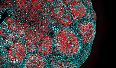
Time-resolved scanning allows the 3D visualization of dynamic structures in a eukaryotic cell membrane at nanometer resolutions.
Studying the functions of living cells and organelles at the nanoscale is vital to understanding the causes of disease. Traditional approaches, including electron microscopy, may, unfortunately, damage these cells.
The Swiss researchers developed a SCIM microscope that resolves spatiotemporally diverse three-dimensional processes on a eukaryotic cell membrane at sub-5 nanometer axial resolution. This may offer insights into intracell interactions in the fight against infections and disease.
The Origins of Scanning Probe Microscopy
Studying the intricate functions of living cells at the nanoscale is a unique challenge. Researchers have developed a range of techniques to meet this challenge, including atomic force microscopy (AFM), scanning tunneling microscopy (STM) and Scanning Probe Electrochemistry (SPE).
Scanning probe microscopy (SPM) forms images of surfaces using a probe that scans the specimen. The technique made its first appearance in 1981 in the form of the scanning tunneling microscope, which produces images at atomic resolution by scanning a specimen using a probe.
In scanning probe microscopes, piezoelectric actuators move the probe with atomic-level precision controlled by electronics. The probe raster scans the specimen. It captures discrete data points which are used to form an image. Its manner of scanning is called a mode.
Scanning Ion Conductance Microscopy (SICM) was developed by P.K. Hansma and his colleagues at the University of California in 1989. An electrolyte-containing aqueous medium is a poor conductor.
A SCIM microscope scans a nanoprobe (micro-pipette with a 50 to 100 nm hole) close to the surface of the specimen. As the probe moves across the specimen, ionic currents flow through the pipette. The strengths of these currents vary according to the electrical resistance across the sample’s surface, thus revealing information about its composition.
In the hopping mode described by the Swiss team, however, the nanoprobe is not raster scanned. It moves vertically up and down in a hopping motion.
The probe approaches the specimen at a distance of 25 to 50 nm at specified points and retracts, thus providing discrete points of measurement from which an image is formed. Crucially, the probe never touches the specimen, thus preventing damage to the sample.
SCIM microscopy is, therefore, a powerful tool for capturing the steep changes in a cell’s topography without affecting the sample.
Time-Resolved Scanning Ion Conductance Microscopy
Time-resolved SICM microscopes produce high-resolution profiles of cell shapes and surface characteristics. However, these need to be correlated with biochemical information and changes to the internal organization of the cells.
The Swiss team integrated an inverted optical microscope to a SICM microscope which allowed them to combine recently developed super-resolution microscopy techniques into their approach.
The SICM setup consisted of a custom-built pipette Z-actuator (vertical actuator) integrated into a controlled-atmosphere device, critical for cell viability during imaging.
The imaging of eukaryotic cells requires piezo actuators with a long-range (>10−20 μm). This leads to a trade-off between resonance frequency and the range of the actuator. The team overcame this by adaptively slowing down the pipette’s velocity and applying a gain to the piezo motion as a function of the current interaction curve.
The Z-actuator achieved a wide mechanical displacement amplification of 22 μm scanning range on the cell surface. It was driven by a custom-made piezo controller and integrated with a stepper-motor stage for approaching the sample.
The team used borosilicate and quartz nanopipettes for probing. They were fabricated with a CO2 laser puller with a radius of 20 to 60 nm in size. Quartz capillaries were shrunk to a sub-10-nm radius using electron beam irradiation.
Many cellular processes occur at time scales of minutes or hours and are easily trackable with time-lapse SICM. Subcellular processes, such as endocytosis or infection, however, occur much faster. The Swiss team’s technique combines the capability to address large imaging volumes (up to 220 000 μm3) relatively quickly with high-speed SICM imaging of small details on live cells.
The wide range of measurements possible (Scan sizes from 500 × 500 nm2 to 100 × 100 μm2, imaging speeds from 0.5 s/image to 20 min/image; Number of pixels per image from 1 Kp to 1 Mp; Depth of view of 22 μm with axial resolution below 10 nm) significantly enhances the range of biological studies facilitated by SICM microscopy.
News

New book from Nanoappsmedical Inc. – Global Health Care Equivalency
A new book by Frank Boehm, NanoappsMedical Inc. Founder. This groundbreaking volume explores the vision of a Global Health Care Equivalency (GHCE) system powered by artificial intelligence and quantum computing technologies, operating on secure [...]

RNA Recycling Extends Lifespan
Summary: Researchers discovered a biological “trash disposal” mechanism that directly controls how fast we age. While circular RNA has long been known to accumulate in cells as we get older, this study proves for the [...]

Cancer’s Deadly Paradox: How Tumors Break Their Own DNA To Keep Growing
Cancer’s strongest gene switches push DNA into damaging overdrive, creating repeated breaks and repairs that may fuel tumor evolution while exposing possible therapeutic weak spots. A new study indicates that cancer can harm its own genetic [...]

NanoMedical Brain/Cloud Interface – Explorations and Implications. A new book from Frank Boehm
New book from Frank Boehm, NanoappsMedical Inc Founder: This book explores the future hypothetical possibility that the cerebral cortex of the human brain might be seamlessly, safely, and securely connected with the Cloud via [...]

Our books now available worldwide!
Online Sellers other than Amazon, Routledge, and IOPP Indigo Global Health Care Equivalency in the Age of Nanotechnology, Nanomedicine and Artifcial Intelligence Global Health Care Equivalency In The Age Of Nanotechnology, Nanomedicine And Artificial [...]
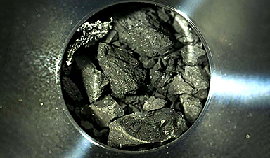
Ryugu asteroid samples contain all DNA and RNA building blocks, bolstering origin-of-life theories
All the essential ingredients to make the DNA and RNA underpinning life on Earth have been discovered in samples collected from the asteroid Ryugu, scientists said Monday. The discovery comes after these building blocks [...]

Is Berberine Really a “Natural Ozempic”?
Often labeled a “natural Ozempic,” berberine is widely discussed as a metabolic aid. Yet research suggests its influence may lie deeper. In recent years, berberine has gained significant attention as a supposed “natural way” [...]
Viagra Ingredient Shows Promise for Rare Childhood Brain Disease in Surprising Study
A rare childhood disease with no approved treatment may have an unexpected new therapeutic candidate. Sildenafil, the active ingredient also sold under the brand name Viagra, may help reduce symptoms in people with Leigh [...]

In a first for China, Neuracle’s implantable brain-computer interface wins approval
In a landmark development, Neuracle Medical Technology has secured the country’s first-ever approval for an implantable brain-computer interface (BCI) system designed to restore hand motor function in patients with spinal cord injuries, in a [...]
A Cambridge Lab Mistake Reveals a Powerful New Way to Modify Drug Molecules
A surprising lab discovery reveals a light-powered way to tweak complex drugs faster, cleaner, and later in development. Researchers at the University of Cambridge have created a new technique for altering complex drug molecules [...]

New book from NanoappsMedical Inc – Molecular Manufacturing: The Future of Nanomedicine
This book explores the revolutionary potential of atomically precise manufacturing technologies to transform global healthcare, as well as practically every other sector across society. This forward-thinking volume examines how envisaged Factory@Home systems might enable the cost-effective [...]

Scientists Discover Simple Saliva Test That Reveals Hidden Diabetes Risk
Researchers have identified a potential new way to assess metabolic health using saliva instead of blood. High insulin levels in the blood, known as hyperinsulinemia, can reveal metabolic problems long before obvious symptoms appear. It is [...]

One Nasal Spray Could Protect Against COVID, Flu, Pneumonia, and More
A single nasal spray vaccine may one day protect against viruses, pneumonia, and even allergies. For decades, scientists have dreamed of creating a universal vaccine capable of protecting against many different pathogens. The idea [...]
New AI Model Predicts Cancer Spread With Incredible Accuracy
Scientists have developed an AI system that analyzes complex gene-expression signatures to estimate the likelihood that a tumor will spread. Why do some tumors spread throughout the body while others remain confined to their [...]

Scientists Discover DNA “Flips” That Supercharge Evolution
In Lake Malawi, hundreds of species of cichlid fish have evolved with astonishing speed, offering scientists a rare opportunity to study how biodiversity arises. Researchers have identified segments of “flipped” DNA that may allow fish to adapt rapidly [...]

Scientists Discover Why Some COVID Survivors Still Can’t Taste Food Years Later
A new study provides the first direct biological evidence explaining why some people continue to experience taste loss long after recovering from COVID-19. Researchers have uncovered specific biological changes in taste buds that could help [...]