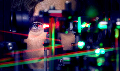
Our ability to see starts with the light-sensitive photoreceptor cells in our eyes. A specific region of the retina, termed fovea, is responsible for sharp vision. Here, the color-sensitive cone photoreceptors allow us to detect even the smallest details. The density of these cells varies from person to person. Additionally, when we fixate on an object, our eyes make subtle, continuous movements, which also differ between individuals.
Researchers from the University Hospital Bonn (UKB) and the University of Bonn have now investigated how sharp vision is linked to these tiny eye movements and the mosaic of cones. Using high-resolution imaging and micro-psychophysics, they demonstrated that eye movements are finely tuned to provide optimal sampling by the cones. The results of the study have now been published in the journal eLife.
Humans can fixate their gaze on an object to see it clearly thanks to a small region in the center of the retina. This area, known as the fovea (Latin for “pit”), is made up of a tightly packed mosaic of light-sensitive cone photoreceptor cells. Their density reaches peaks of more than 200,000 cones per square millimeter—in an area about 200 times smaller than a quarter-dollar coin. The tiny foveal cones sample the portion of visual space visible to the eye and send their signals to the brain. This is analogous to the pixels of a camera sensor with millions of photo‑sensitive cells spread across its surface.
However, there is an important difference. Unlike the pixels of a camera sensor, the cones in the fovea are not uniformly distributed. Each eye has a unique density pattern in their fovea.
Additionally, “unlike a camera, our eyes are constantly and unconsciously in motion,” explains Dr. Wolf Harmening, head of the AOVision Laboratory at the Department of Ophthalmology at UKB and a member of the Transdisciplinary Research Area (TRA) “Life & Health” at the University of Bonn.
This happens even when we are looking steadily at a stationary object. These fixational eye movements convey fine spatial details by introducing ever-changing photoreceptor signals, which must be decoded by the brain. It is well known that one of the components of fixational eye movements, termed drift, can differ between individuals, and that larger eye movements can impair vision. How drift relates to the photoreceptors in the fovea, however, and our ability to resolve fine detail has not been investigated until now.
, AOVision Laboratory / Wolf Harmening")
Using high-resolution imaging and micro-psychophysics
This is precisely what Harmening’s research team has now investigated by using an adaptive optics scanning light ophthalmoscope (AOSLO), the only one of its kind in Germany. Given the exceptional precision offered by this instrument, the researchers were able to examine the direct relationship between cone density in the fovea and the smallest details we can resolve.
At the same time, they recorded the tiny movements of the eyes. To do this, they measured the visual acuity of 16 healthy participants while performing a visually demanding task. The team tracked the path of the visual stimuli on the retina to later determine which photoreceptor cells contributed to vision in each participant. The researchers—including first author Jenny Witten from the Department of Ophthalmology at UKB, who is also a Ph.D. student at the University of Bonn—used AOSLO video recordings to analyze how the participants’ eyes moved during a letter discrimination task.
Eye movements are finely tuned to cone density
The study revealed that humans are able to perceive finer details than the cone density in the fovea would suggest.
“From this, we conclude that the spatial arrangement of foveal cones only partially predicts resolution acuity,” reports Harmening. In addition, the researchers found that tiny eye movements influence sharp vision: during fixation, drift eye movements are precisely aligned to systematically move the retina synchronized with the structure of the fovea.
“The drift movements repeatedly brought visual stimuli into the region where cone density was highest,” explains Witten. Overall, the results showed that within just a few hundred milliseconds, drift behavior adjusted to retinal areas with higher cone density, improving sharp vision. The length and direction of these drift movements played a key role.
According to Harmening and his team, these findings provide new insights into the fundamental relationship between eye physiology and vision: “Understanding how the eye moves optimally to achieve sharp vision can help us to better understand ophthalmological and neuropsychological disorders, and to improve technological solutions designed to mimic or restore human vision, such as retinal implants.”
More information: Sub-cone visual resolution by active, adaptive sampling in the human foveolar, eLife (2024). DOI: 10.7554/eLife.98648.3
News

Scientists Just Discovered the Hidden Trick That Keeps Your Cells Alive
A strange bead-like motion inside cells may be the secret to keeping their DNA—and health—in balance. Mitochondria are often described as the power plants of the cell because they produce the energy cells need [...]

Scientists Discover Stem Cells That Could Regrow Teeth and Bone
Scientists just uncovered the cellular “blueprint” that could one day let us regrow real teeth. Researchers at Science Tokyo have uncovered two distinct stem cell lineages that play a central role in forming tooth [...]

Scientists Uncover Fatal Weakness in “Zombie Cells” Linked to Cancer
A newly identified weakness in “zombie” cells may open the door to more precise cancer treatments by turning their own survival strategy against them. A new class of drugs takes advantage of a recently [...]

Bowel and Ovarian Cancers Are Dramatically Rising in Young Adults, Scientists Aren’t Sure Why
Cancer incidence is increasing, especially among younger adults, and current risk factors don’t fully account for the trend. Scientists suggest other underlying causes may be contributing. Cancer patterns in England are shifting in a [...]

New Immune Pathway Could Supercharge mRNA Cancer Vaccines
A surprising backup system in the immune response to mRNA vaccines may hold the key to more effective cancer treatments. The arrival of mRNA vaccines against SARS-CoV-2 in 2020 marked a turning point in the COVID-19 pandemic. Today, [...]
Scientists Discover “Molecular Switch” That Fuels Alzheimer’s Brain Inflammation
A newly identified trigger of brain inflammation could offer a fresh target for slowing Alzheimer’s progression. The brain has its own built-in immune system that identifies threats and responds to them. In Alzheimer’s disease, growing evidence [...]

Molecular Manufacturing: The Future of Nanomedicine – New book from NanoappsMedical Inc.
This book explores the revolutionary potential of atomically precise manufacturing technologies to transform global healthcare, as well as practically every other sector across society. This forward-thinking volume examines how envisaged Factory@Home systems might enable the cost-effective [...]

Forgotten Medicinal Plant Shows Promise in Fighting Dangerous Superbugs
A traditional medicinal plant, tormentil, shows promise against antibiotic-resistant bacteria in laboratory tests. Its compounds work by limiting bacterial growth and boosting antibiotic performance. Before the development of modern antibiotics, plant-based remedies were commonly [...]

NanoMedical Brain/Cloud Interface – Explorations and Implications. A new book from Frank Boehm
New book from Frank Boehm, NanoappsMedical Inc Founder: This book explores the future hypothetical possibility that the cerebral cortex of the human brain might be seamlessly, safely, and securely connected with the Cloud via [...]

New Research Finds Shocking Link Between Chili Peppers and Cancer
If you love spicy food, you are not alone. But scientists are taking a closer look at whether eating a lot of chili peppers could affect your cancer risk. Could your love of spicy [...]

New book from Nanoappsmedical Inc. – Global Health Care Equivalency
A new book by Frank Boehm, NanoappsMedical Inc. Founder. This groundbreaking volume explores the vision of a Global Health Care Equivalency (GHCE) system powered by artificial intelligence and quantum computing technologies, operating on secure [...]
Scientists Create “Neurobots” – Living Machines With Their Own Nervous Systems
Neurobots—xenobots with neurons—show self-organized nervous systems and enhanced behaviors, revealing new insights into how biology builds functional structures. In 2020, researchers at Tufts University developed tiny living structures known as xenobots using frog cells. These microscopic organisms [...]

Our books now available worldwide!
Online Sellers other than Amazon, Routledge, and IOPP Indigo Global Health Care Equivalency in the Age of Nanotechnology, Nanomedicine and Artifcial Intelligence Global Health Care Equivalency In The Age Of Nanotechnology, Nanomedicine And Artificial [...]

Amazonian Chocolate Could Become the Next Superfood, Scientists Say
New research into Amazonian cocoa reveals that its value may extend beyond flavor alone. Chocolate from the Amazon is already known worldwide for its distinctive taste, but new research suggests it may offer even [...]
Nanobody repairs misfolded CFTR inside cells, boosting function in cystic fibrosis
A tiny antibody component could fundamentally transform the treatment of cystic fibrosis: For the first time, researchers have succeeded in developing a so-called nanobody that penetrates directly into human cells and can repair the [...]

20-Year Study Finds Daily Multivitamins Don’t Extend Lifespan
A large, decades-long study of over 390,000 U.S. adults challenges a widespread assumption about daily multivitamins. Multivitamins are a daily habit for millions of Americans, often taken with the expectation that they will extend [...]