Doxorubicin (DOX) is a powerful anti-cancer medication, and efforts have been made to design nanostructures for delivering it to cancerous cells. The nanostructures increase the cytotoxic effects of DOX on cancerous cells, while reducing the negative effects on healthy cells.
In a research paper published in the journal Scientific Reports, layered double hydroxide (LDH) nanostructures were developed to administer DOX effectively.
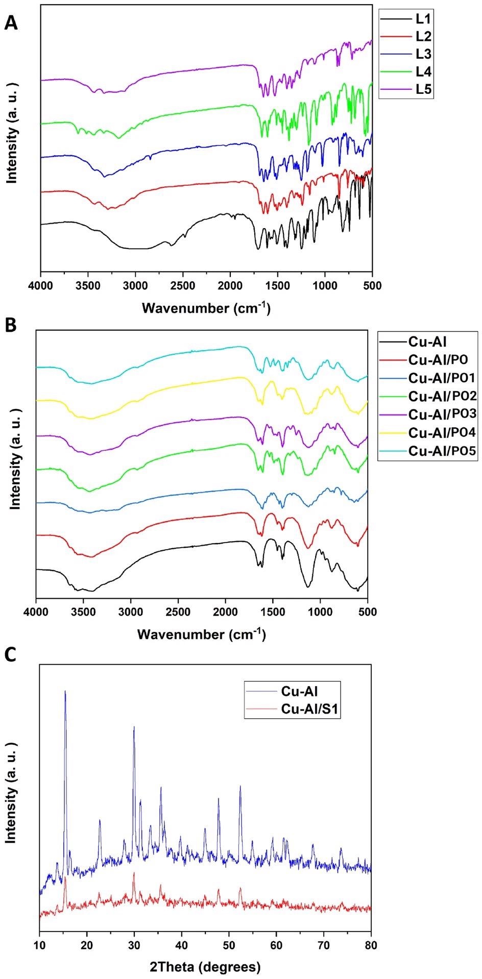
Figure 1. FTIR spectra of the synthesized nanomaterials (A,B). XRD results of the synthesized nanomaterials (C). S1 in part (C) stands for one of the PO’s. © Kiani, M., Bagherzadeh, M., Ghadiri, A. M., Makvandi, P., & Rabiee, N. (2022).
Cancer, and How Nanotechnology Can Help
Cancer accounts for the second-largest mortality rate after cardiac illnesses, with susceptibility to tumor development determined by multiple factors like age, family history and carcinogen exposure.
Chemotherapy is the most often used cancer treatment. However, complex encounters in the cancer microenvironment, as well as cancer cells’ capacity to proliferate and flip between molecular routes to guarantee their preservation, have led to the tumors developing resistance against treatments.
Doxorubicin (DOX) is widely used in tumor treatment because it may limit DNA replication by decreasing the action of topoisomerase enzymes, suppressing cell cycle progress and ultimately directing tumors towards cellular death.
Resistance against DOX has been caused by a range of factors, including discharge of doxorubicin by P-glycoprotein from tumor cells, Bcl-2 overexpression, apoptotic suppression, and abnormal expression of epigenetic and genetic variables. As a result, research has concentrated on developing nanostructured delivery mechanisms for doxorubicin to increase its cancer-suppressing effectiveness.
Benefits of LDH as Drug Deliver Systems
Nanotechnology offers fresh promise for reducing resistance against treatments and improving the efficacy of chemotherapy drugs in cancer treatment.
Layered double hydroxides (LDHs) are multilayer nanoscale structures and anion clays having a hydrotalcite crystalline structure generated by two metallic ions, comprising a trivalent and a divalent metallic ion, an -OH group, an H2O molecule, and an interlayer anion.
Due to their distinctive multilayer architecture and interlayer anion exchanging capability, LDH nanostructures have paved the way in the biomedicinal domain. One of the most significant uses of LDH nanostructures in the administration of drugs is the incorporation of a specific chemical into LDHs through an interlayer anionic exchange.
LDHs have excellent drug loading capability, a large surface area, great durability, and anion exchanging ability. These properties make them preferable for drug administration, especially when compared to other nanostructures like polymer nanoparticles (NPs).
More significantly, as LDHs have a dissoluble bulk layer at pH 5.0 (around the acidity level of the cancer microenvironment), they are great contenders for drug administration against tumors.
Advantages of Plantago Ovata
Plantago ovata (PO) is a classic botanical remedy with bioactive polysaccharides. It is an organically produced substance with advantages such as sustainable production, low cost, availability, and a good safety profile.
PO was initially employed to treat wounds. Subsequent research revealed that PO extracts possess a variety of medicinal properties, such as antioxidant, anti-inflammation, immunomodulation, and pain-relieving properties.
Important Findings of the Study
PO was utilized to modify the surfaces of Cu–Al LDH nanoscale structures to boost their capability as nanoscale drug administration systems. Doxorubicin, an anti-cancer medication, was stacked onto NPs once they were prepared, and characterization procedures showed proper fabrication and drug content.
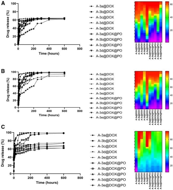
The drug discharge analysis indicated pH-sensitive discharge of doxorubicin from LDH NPs, with the largest discharge of anti-cancer medications occurring at pH 5.5 and the least amount of medication discharge occurring at pH 4.5, perhaps owing to the detrimental effect of lower and strongly acidic pH on NP architectures.
The MTT experiment exhibited great cytocompatibility of PO-incorporated Cu–Al LDH nanostructures, demonstrating partial and low cytotoxicity against HEK-293 and PC12 cells, while decreasing the viability of MCF-7 and HT-29 cells as cancerous cells.
Notably, the decrease in viability of HT-29 and MCF-7 cells was smaller in PO-incorporated LDH NPs than LDH nanoscale carriers without PO, which should be investigated further in future studies. The CLSM data demonstrated that LDH NPs delivered doxorubicin to the nucleus and cytoplasm of HEK-293 and MCF-7 cells.
Histological examination of renal tissue revealed no cellular deterioration, adequate cellular and tubular architecture, and zero bloodstream obstructions. This indicates the great cytocompatibility of PO-incorporated LDH nanostructures.
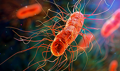
Antimicrobial testing revealed that Cu–Al LDH nanoparticles exhibited biotoxicity against Gram-negative and Gram-positive bacteria, and they may be used in treating microbial illnesses in future trials.
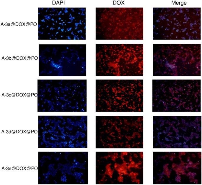
Figure 3. The CLSM images of the drug loaded nanocarriers-coated with leaf extracts treated with HEK-293 cell lines. The used concentration of the nanoparticles: 17.5 μg/mL. © Kiani, M., Bagherzadeh, M., Ghadiri, A. M., Makvandi, P., & Rabiee, N. (2022).
News
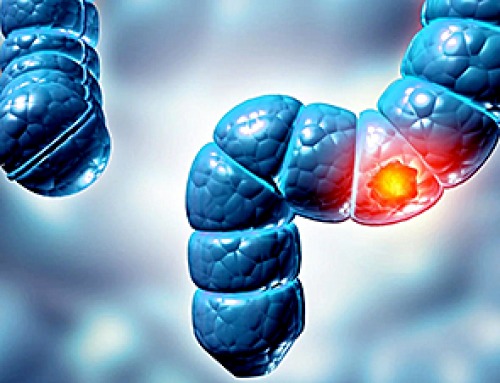
Why More People in Their 30s Are Suddenly Getting Colon Cancer
A major Swiss study found that colorectal cancer is becoming increasingly common in adults under 50, even as rates decline in older age groups. Researchers in Switzerland have identified a concerning trend: while colorectal [...]
Researchers Compare MS Models to Human Tissue in Search for Better Therapies
Researchers identified key differences between two widely used multiple sclerosis models, showing how each can better study myelin damage, immune responses, and repair. The findings may improve efforts to develop treatments that restore lost [...]
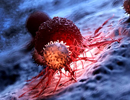
Scientists Discover Genetic “Off Switch” That Supercharges CAR T Cells Against Cancer
A new study reveals a possible way to make CAR T-cell therapy more durable and effective by targeting a single gene-regulating protein. CAR T-cell therapy is widely seen as a breakthrough in personalized cancer [...]

New Vitamin B12-Based Therapy Could Change How Brain Cancer Is Treated
Researchers have identified a vitamin B12–based compound that appears capable of crossing the blood–brain barrier and selectively accumulating in glioblastoma tissue. For decades, one of the biggest problems in brain cancer treatment has had [...]

Simple Fiber Supplement Cuts Knee Arthritis Pain in Just 6 Weeks, Study Finds
A daily inulin supplement may help reduce knee osteoarthritis pain while revealing a possible link between gut health, muscle function, and pain sensitivity. For millions of people living with knee osteoarthritis, managing chronic pain [...]

This Common Vitamin May Help Stop Prediabetes From Turning Into Diabetes
Vitamin D may help prevent type 2 diabetes in people with specific genetic variations, offering a possible path toward personalized diabetes prevention. More than 40% of U.S. adults have prediabetes, a condition in which [...]

Ebola, hantavirus: Is the world prepared for the next pandemic?
Funding cuts to health research and a growing antivaccine movement are making it harder than ever to respond to viruses. The World Health Organization (WHO) has declared that an Ebola outbreak in Uganda and [...]

May 2026 Healthcare News and Trends: Market Signals That Matter
Artificial intelligence is dominating headlines, telehealth has settled into a new normal, and digital health continues to promise transformation. However, much of what is being discussed in healthcare today reflects potential rather than reality. [...]
Scientists Rewire Donor Stem Cells To Outsmart Aggressive Blood Cancers
Researchers have tested a gene-edited stem cell transplant designed to shield healthy blood-forming cells from powerful cancer-targeting immunotherapies. For patients with highly aggressive blood cancers, stem cell transplantation can offer a rare chance at [...]

Recent Digital Health Trends, Insights and News – May 2026
Last month marked continued progress as digital health moves into its next phase — from AI expanding into drug discovery and core infrastructure to new federal pathways accelerating device access and home-based care. Together, [...]
Cancer Mystery Solved: Scientists Discover How Melanoma Becomes “Immortal”
Scientists have uncovered a previously overlooked mechanism that may help melanoma cells become effectively “immortal.” Cancer cells face a major problem before they can become deadly: They have to figure out how to stop [...]
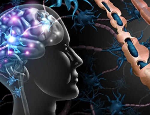
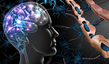
How Visual Neurons Organize Thousands of Synaptic Inputs
Summary: A new study uncovered the organizational rules that determine how neurons in the primary visual cortex process information. By imaging both the cell bodies (soma) and the individual synapses (on dendritic spines) of [...]

Scientists Just Found a Surprising Way To Destroy “Forever Chemicals”
Scientists have uncovered a new mechanism that may help break down highly persistent PFAS pollutants. PFAS have earned the nickname “forever chemicals” for a reason. These industrial compounds are so chemically durable that they [...]
Scientists Discover Cheap Material That Kills Deadly Superbugs
A new sulfur-rich antimicrobial polymer shows strong effectiveness against fungal and bacterial pathogens and may offer an affordable solution to antimicrobial resistance. Antimicrobial resistance is creating growing challenges for both healthcare and food production, [...]

What to Know About Cicada, or BA.3.2, the Latest SARS-CoV-2 Variant Under Monitoring
Like periodical cicadas, the insects for which it is nicknamed, SARS-CoV-2 Omicron subvariant BA.3.2 is only just beginning to emerge after lying low for an extended period since it first appeared. Although it was [...]
Scientists Say This Simple Supplement May Actually Reverse Heart Disease
Scientists in Japan say a common supplement may actually help “unclog” certain diseased heart arteries from the inside out. A simple food supplement sold in Japan may have helped reverse a dangerous form of [...]