Unprecedented views of the interior of cells and other nanoscale structures are now possible thanks to innovations in expansion microscopy. The advancements could help provide future insight into neuroscience, pathology, and many other biological and medical fields.
In the paper “Magnify is a universal molecular anchoring strategy for expansion microscopy,” published today (January 2, 2023) in the journal Nature Biotechnology, collaborators from Carnegie Mellon University, the University of Pittsburgh, and Brown University describe new protocols for dubbed Magnify.
“Magnify can be a potent and accessible tool for the biotechnology community,” said Yongxin (Leon) Zhao, the Eberly Family Career Development Associate Professor of Biological Sciences.
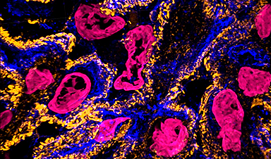
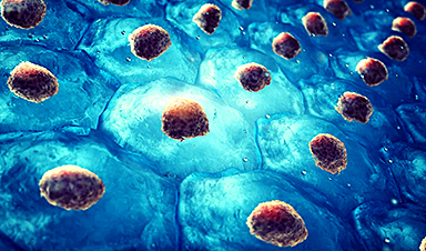
A video shows kidney cells. Expansion microscopy (ExM) provides unprecedented views of cell interiors. The emerging super-resolution imaging technique relies on physical — rather than optical — magnification. Advancements by Carnegie Mellon University’s Zhao Biophotonics Lab increases the expansion rate and allows many types of tissues to be viewed in 3D. Credit: Carnegie Mellon University
Magnify is a variant of expansion microscopy that allows researchers to use a new hydrogel formula, invented by Zhao’s team, that retains a spectrum of biomolecules, offers a broader application to a variety of tissues, and increases the expansion rate up to 11 times linearly or ~1,300 folds of the original volume.
“We overcame some of the longstanding challenges of expansion microscopy,” Zhao said. “One of the main selling points for Magnify is the universal strategy to keep the tissue’s biomolecules, including proteins, nucleus snippets, and carbohydrates, within the expanded sample.”
Zhao said that keeping different biological components intact matters because previous protocols required eliminating many various biomolecules that held tissues together. But these molecules could contain valuable information for researchers.
“In the past, to make cells really expandable, you need to use enzymes to digest proteins, so in the end, you had an empty gel with labels that indicate the location of the protein of interest,” he said. With the new method, the molecules are kept intact, and multiple types of biomolecules can be labeled in a single sample.
“Before, it was like having single-choice questions. If you want to label proteins, that would be the version one protocol. If you want to label nuclei, then that would be a different version,” Zhao said. “If you wanted to do simultaneous imaging, it was difficult. Now with Magnify, you can pick multiple items to label, such as proteins, lipids, and carbohydrates, and image them together.”
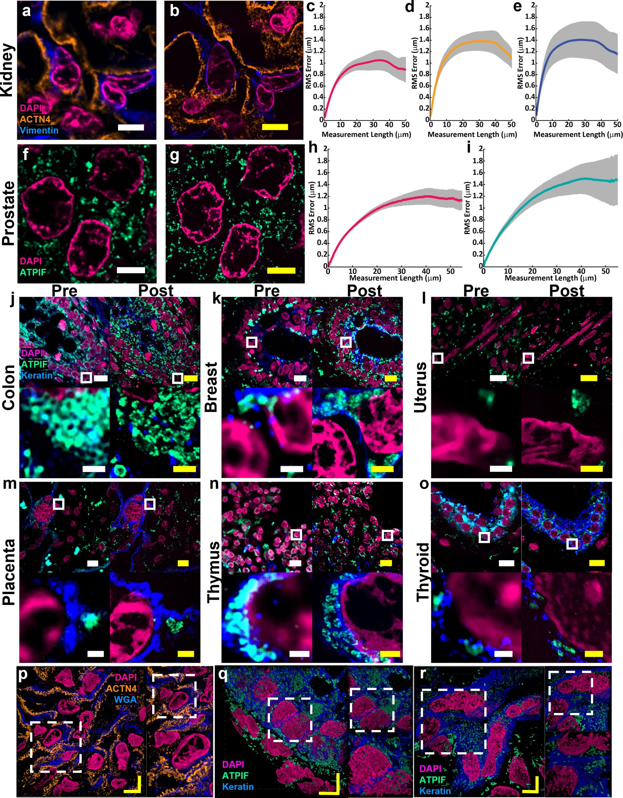
Example of (a) pre-expansion images of human kidney imaged at 60× and processed with SOFI compared to the same field of view (b) post-expansion with MAGNIFY taken at 40×. Magenta, DAPI; Orange, anti-alpha-actinin 4 (ACTN4); Blue, vimentin. Post expansion images are maximum intensity projected over 25 frames in z. (c-e) Root mean square (RMS) length measurement error as a function of measurement length for pre-expansion versus post expansion images for (c) DAPI, (d) ACTN4, and (e) Vimentin. Solid line, mean of channel; shaded area, standard error of mean (s.e.m); n = 5 technical replicates; average expansion factor, 8.64× (s.e.m 0.24). Example of (f) pre-expansion images of human prostate imaged at 60× and processed with SOFI compared to the same field of view (g) post-expansion with MAGNIFY taken at 40×. Magenta, DAPI; Green, Anti-ATPase Inhibitory Factor 1 (ATPIF). Post expansion images maximum intensity projected over 3 frames. (h-i) RMS length measurement error as a function of measurement length for pre-expansion versus post expansion images of (h) DAPI, and (i) ATPIF. Solid line, mean of channel; shaded area, s.e.m.; n = 4 technical replicates; average expansion factor, 10.38× (s.e.m 0.57). (j-o) Validation of MAGNIFY across multiple human tissue types. FFPE samples of human tissue were imaged at 40× (top left). Images were taken at 60×and processed with SOFI (bottom left). The white box indicates the field of view of the higher magnification images. The samples were then processed with the MAGNIFY protocol, and the same fields of view were imaged post-expansion in water at 10× (top right) and 40× (bottom right). Post expansion images were projected over 4-17 z slices. Magenta, DAPI; Green, ATPIF; Blue, Cytokeratin Pan Type I/II. Expansion factors in water were (j) Colon: 8.85×, (k) Breast: 9×, (l) Uterus: 8×, (m) Placenta: 8.75×, (n) Thymus: 10.00×, (o) Thyroid: 10.59×. (p-r) Example 3d images of human tissues: (p) kidney (Expansion factor 8.68×). Magenta, DAPI; Orange, ACTN4; Blue, WGA. (q) colon (Expansion factor 9.67×). Magenta, DAPI; Green, ATIPF; Blue, Cytokeratin Pan Type I/II. (r) Uterus (Expansion factor 8×). Magenta, DAPI; Green, ATIPF; Blue, Cytokeratin Pan Type I/II. Zoomed in regions indicated by dashed white box. Scale bars (yellow indicates post expansion images): (a) 5 μm; (b) 5 μm (physical scale post expansion: 40.75 μm; expansion factor: 8.15×); (f) 5 μm; (g) 5 μm (physical scale post expansion: 51.9 μm; expansion factor: 10.38×); (j-o) top: 10 μm; bottom: 1 μm; (p-t) 5 μm. Scale bars are all in biological scale. Credit: Courtesy of Carnegie Mellon University
Lab researchers Aleksandra Klimas, a postdoctoral researcher and Brendan Gallagher, a doctoral student, were first co-authors on the paper.
“This is an accessible way to image specimens in high resolution,” Klimas said. “Traditionally, you need expensive equipment and specific reagents and training. However, this method is broadly applicable to many types of sample preparations and can be viewed with standard microscopes that you would have in a biology laboratory.”
Gallagher, who has a background in neuroscience, said their goal was to make the protocols as compatible as possible for researchers who could benefit from adopting the Magnify as part of their toolkits.
“One of the key concepts that we tried to keep in mind was to meet researchers where they are and have them change as few things in their protocols as possible,” Gallagher said. “It works with different tissue types, fixation methods and even tissue that has been preserved and stored. It is very flexible, in that you don’t necessarily need to redesign experiments with Magnify in mind completely; it will work with what you have already.”
For researchers such as Simon Watkins, the founder and director of the Center for Biologic Imaging at the University of Pittsburgh and the Pittsburgh Cancer Institute, the fact that the new protocol is compatible with a broad range of tissue types — including preserved tissue sections — is important. For example, most expansion microscopy methods are optimized for brain tissue. In contrast, Magnify was tested on samples from various human organs and corresponding tumors including breast, brain and colon.
“Let’s say you have a tissue with dense and non-dense components, this gets around tissues that previously wouldn’t expand isometrically,” Watkins said. “Leon has been working hard on this to make this protocol work with tissues that have been archived.”
Xi (Charlie) Ren, an assistant professor of biomedical engineering at Carnegie Mellon, studies the lung tissue and how to model its morphogenesis and pathogenesis. Part of his research involves researching the motile cilia that function to clear mucus in the human conducting airway. At 200 nanometers in diameter and just a few micrometers in length, the structures are too small to see without time-intensive technology such as electron microscopy. Working in collaboration with Zhao’s lab, Ren’s team developed and delivered lung organoid models with specific defects in cilia ultrastructure and function to validate the ability of Magnify to visualize clinically relevant cilia pathology.
“With the latest Magnify techniques, we can expand those lung tissues and start to see some ultrastructure of the motile cilia even with a regular microscope, and this will expedite both basic and clinical investigations,” he said.
The researchers also were able to view defects in cilia in patient-specific lung cells known to have genetic mutations.
“The lung tissue engineering community always needs a better way to characterize the tissue system that we work with,” Ren said. He added that this work is an important first step and he hopes the collaborative work with Zhao’s lab will further be refined and applied to pathology samples found in tissue banks.
Finally, the hydrogel used in Magnify and developed in the Zhao lab is more robust than its predecessor, which was very fragile, causing breaks during the process.
“We are hoping to develop this technology to make it more accessible to the community,” he said. “There are different directions this can go. There’s a lot of interest in using this kind of tissue expansion technology for basic science.”
Alison Barth, the Maxwell H. and Gloria C. Connan Professor in the Life Sciences at Carnegie Mellon, studies synaptic connectivity during learning. She said the broad applications provided by the new methods will be a boon for researchers.
“The brain is a great place to take advantage of these super-resolution techniques,” said Barth, who collaborates with the Zhao Lab on several studies. “Microscopy methods will be beneficial for synaptic phenotyping and analysis across different brain conditions.
“One of the major advances in this paper is the method’s ability to work on many different types of tissue specimens.”
News

How Immune Dysregulation Contributes to Psychiatric Disorders
Introduction Growing evidence suggests that disruptions in immune function may play an important role in the development and progression of psychiatric disorders. However, immune mechanisms probably contribute more strongly in some patients than others, [...]

Electrostatic Discharge Boosts Triboelectric Nanogenerator Current and Enables DC Output
Controlled electrical discharges could enable triboelectric nanogenerators to achieve higher peak currents, extending nano-enabled energy harvesting into chemical processing and self-powered sensing. Paper: Electrostatic discharge as a breakthrough strategy for triboelectric nanogenerators. A new review [...]
Swiss laboratory uses old drugs against rare diseases
Researchers at the University of Geneva are combing through collections of approved drugs to find new therapies for rare diseases – with some success. This approach is gaining traction around the world, while pharmaceutical [...]
Nanozyme Aptasensors Show Promise for Faster Food, Health, and Environmental Testing
By pairing robust artificial enzymes with highly selective aptamers, nanozyme aptasensors could help detect disease biomarkers, pathogens, and contaminants faster, but the review shows that real-world deployment still depends on overcoming matrix interference, biofouling, [...]

Paralyzed Man Feels Sensation Again With Brain Stimulation Device
Aneuroprosthetic system has allowed a man with paralysis to grasp and lift objects and feel touch again. The device helped 42-year-old Keith Thomas of Massapequa, New York, who was paralyzed from the chest down [...]
Global Cancer Cases Could Surge 67% by 2050, New Report Warns
New data reveal major geographic disparities and highlight the urgent need for global action on prevention, early detection, and equitable access to treatment. For roughly one in five people worldwide, cancer will become part [...]
A Deadly Ebola-Like Virus Is Spreading. Are We Ready?
BU virologist Nancy Sullivan says the Bundibugyo outbreak in the Democratic Republic of the Congo underscores the need for broader outbreak preparedness. The death of a nurse marked the moment health officials recognized that [...]

Why Most Animal Viruses Never Become Human Pandemics
From receptor mismatch to risky human-animal interfaces, this article explains why spillover is common but true pandemic emergence remains rare. Introduction Humans are constantly exposed to animal viruses through farming, wildlife contact, and the [...]

Stem cell organoids repair heart microvessels in coronary artery disease models
A Stanford University team has shown that vascular organoids derived from human stem cells can repair the heart’s microvessel network in pigs with ischaemic heart disease – a proof-of-concept advancement that could open new therapeutic [...]

Goodbye GP waiting rooms, hello prevention at home
Prevention is suddenly everywhere in NHS reform. The recent £340m community pharmacy deal is moving more services onto the high street. Community Diagnostic Centres are being expanded, and the Neighbourhood Health Framework sets out [...]
Ebola control is weakened by mistrust and cultural insensitivity
Effective response depends on cooperation with communities and frontline workers, writes Zaeem ul Haq The current Bundibugyo Ebola outbreak in the Democratic Republic of the Congo (DRC) and Uganda is exposing dangerous gaps in [...]

Building the Brain Requires Millions of Dangerous DNA Breaks
Scientists discovered that building a healthy brain involves an unexpected step: young neurons routinely break and rapidly repair their own DNA. As the brain develops, newly formed nerve cells must travel through tightly packed tissue [...]

One Tiny Change May Explain How Viruses Jump From Bats to Humans
Scientists found that one tiny genetic change may determine whether a bat virus stays in bats or becomes a human threat. Most infectious disease outbreaks begin when a virus or other pathogen crosses from animals into [...]

Scientists Discover 250+ Genes That Could Lead to New Ways To Prevent Melanoma
The world’s largest study of mole genetics identified hundreds of genes tied to melanoma risk, uncovering potential new drug targets and paving the way for more accurate melanoma screening and prevention. Researchers at QIMR [...]
Breakthrough Diabetes Treatment Reprograms the Immune System
An engineered stem cell therapy reversed new-onset Type 1 diabetes in mice by shifting the immune system away from attacking insulin-producing cells. For more than a century, people with Type 1 diabetes have relied [...]
Taking the world’s temperature: WHO chief spotlights global health emergencies
Taking the world’s temperature on pressing health matters, WHO Director-General Tedros Adhanom Ghebreyesus provided the latest on current global challenges - and successes when it comes to international cooperation. “The outbreaks of hantavirus, Ebola and Marburg all show [...]