Researchers identify a shared RNA-protein interaction that could lead to broad-spectrum antiviral treatments for enteroviruses.
A new study from the University of Maryland, Baltimore County (UMBC), published in Nature Communications, explains how enteroviruses begin reproducing inside human cells. These viruses include those responsible for polio, encephalitis, myocarditis, and the common cold, and they start infection by taking over the cell's own molecular machinery.
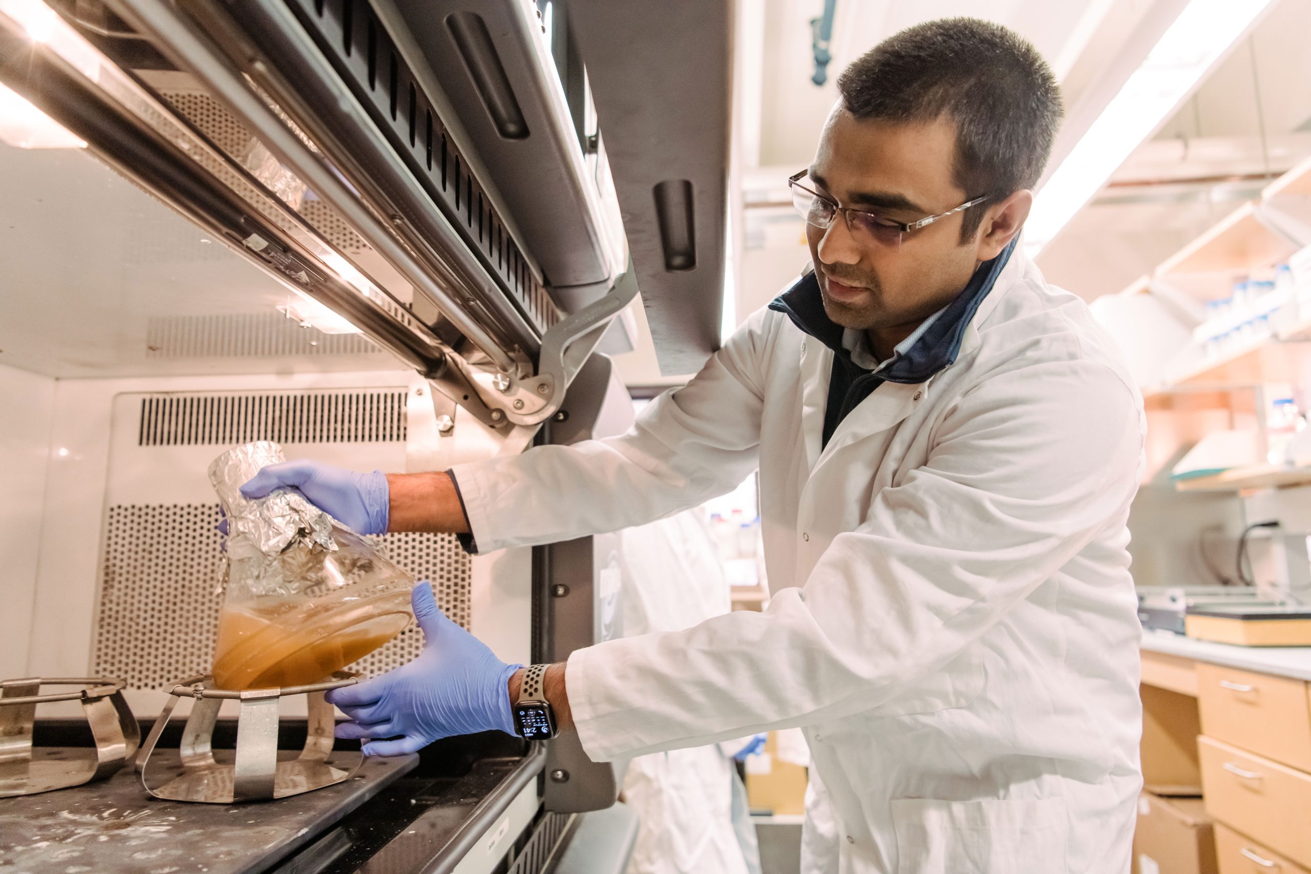
The research was led by senior author Deepak Koirala, associate professor of chemistry and biochemistry, along with recent Ph.D. graduate Naba Krishna Das, and it addresses a long-standing gap in understanding this early stage of viral replication. The findings could also help guide the development of antiviral drugs that work against multiple enteroviruses.
"My lab has been really motivated to understand how RNA viruses produce their proteins inside the cell and multiply their genome to make more virus particles," Koirala says. Building on their discovery of a crucial cloverleaf structure in the viral RNA, Koirala's group has now shown how it recruits proteins to assemble the replication complex.
Seeing the bigger picture
Enteroviruses carry a very small RNA genome that must perform two essential tasks. It serves as instructions for making viral proteins and also acts as the template for copying itself to create new virus particles. While most of this compact genome codes for structural components, it also produces a limited set of proteins that are crucial for replication and not found in human cells.
One of these proteins is a combined molecule known as 3CD. One portion (3C) functions like molecular scissors, cutting the long chain of amino acids produced from the viral RNA into separate working proteins. The other portion (3D) acts as an RNA polymerase, the enzyme responsible for copying the viral RNA. Because human cells do not contain this type of polymerase, the virus must supply it on its own.
"We previously determined the structure of the RNA alone, and other groups determined the structure of 3C and 3D, but now we've captured the structure of the RNA and proteins together, so we know how they are interacting," Koirala explains. "We found that it's the 3C domain of 3CD that binds to the RNA in the viral genome, and then it recruits the other components, such as host protein PCBP2, to assemble the replication complex."
The same complex also works as an on-off switch: when 3CD is attached, the virus copies its RNA; when it lets go, the RNA can be read to make proteins instead.
Resolving a debate
Koirala's team used X-ray crystallography to visualize the interactions between the RNA cloverleaf and 3CD. They augmented those observations with isothermal titration calorimetry (ITC), a technique that quantifies the strength of an interaction by measuring the heat released when molecules bind, and biolayer interferometry (BLI), which tracks light interference to gauge binding duration.
The team also settled a debate by showing that two complete 3CD molecules (bringing two RNA polymerases) bind side-by-side on the RNA, rather than forming a single fused pair, as research from another group had suggested. Why two are needed is still a mystery, but the picture is now clear.
New therapeutic targets
Perhaps most exciting, the seven types of enteroviruses the paper investigated all employed a very similar binding mechanism and RNA cloverleaf structure. The extent of this conservation implies the RNA cloverleaf is very important for replication, and any mutations would likely derail it. That means the RNA and RNA-protein interface is likely to be stable over time and across enteroviruses, making it an even more promising drug target—and opening the door to the tantalizing prospect of a "universal" drug targeting all enteroviruses.
Drugs disrupting 3C and 3D activity are already in development, but "now we have another layer to test," Koirala says. "What if we target the RNA, or the RNA-protein interface, so that we break the interaction? That is another opportunity. Now that we have high-resolution structures, you can precisely design drug molecules to target them."
"Viruses are so, so clever. Their entire genome is equivalent to about one mRNA sequence in humans, yet they are so effective," Koirala says. His latest work demonstrates "why we need to investigate this basic science—so that it can be translated into developing drugs targeting pathogens that cause so many harmful diseases."
Reference: "Structural basis for 3C and 3CD recruitment by enteroviral genomes during negative-strand RNA synthesis" by Naba Krishna Das, Alisha Patel, Reem Abdelghani and Deepak Koirala, 21 October 2025, Nature Communications.
DOI: 10.1038/s41467-025-64376-0
Funding: U.S. National Science Foundation, NIH/National Institutes of Health
News
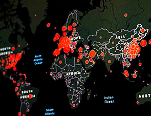
The Corona variant Cicada is here – we know that
Online and on social media, reports are piling up about a new Sars-Cov-2 variant that is currently on the rise: BA.3.2, also known as Cicada. That's what it's all about: The Omicron variant BA.3.2, [...]
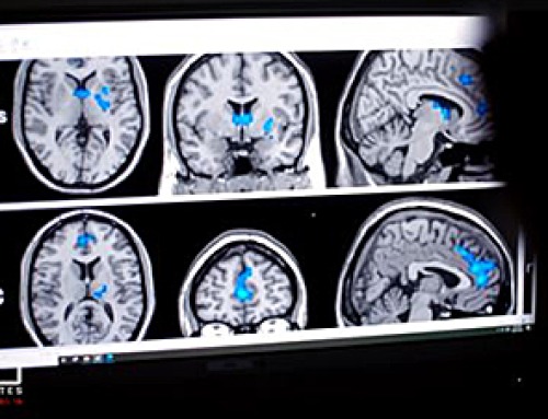
A Simple Blood Test Could Predict Dementia Risk 25 Years Early
A single blood marker may quietly signal dementia risk decades in advance. Scientists at the University of California, San Diego, have identified a blood signal that could forecast dementia risk decades before symptoms begin. Their [...]
Sperm Get Lost in Space and Scientists Finally Know Why
Having a baby in space may be far more complicated than expected, as new research shows sperm struggle to find their way in microgravity. Starting a family beyond Earth could be more complicated than [...]
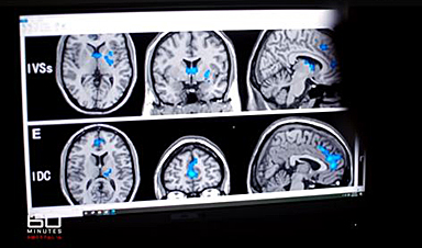
Digital Dementia – Brain fog and disassociation from being chronically online
New medical evidence, featured on 60 Minutes Australia, indicates excessive screen time is causing "digital dementia" in young Australians, with brain scans showing physical shrinkage and damage. Experts warn that high device usage (6-8 hours [...]
A new, highly mutated COVID variant called ‘Cicada’ is spreading in the US.
BA.3.2, a heavily mutated new COVID-19 variant which may be better able to escape immunity from vaccines or prior infection, is now spreading in the United States. Although COVID cases are currently low nationally, [...]

Molecular Manufacturing: The Future of Nanomedicine – New book from NanoappsMedical Inc.
This book explores the revolutionary potential of atomically precise manufacturing technologies to transform global healthcare, as well as practically every other sector across society. This forward-thinking volume examines how envisaged Factory@Home systems might enable the cost-effective [...]

Ancient bacteria strain discovered in ice cave is resistant to some modern antibiotics
In the depths of Scarisoara cave in Romania sits one of the world’s biggest underground glaciers, a monumental slab of ice the size of roughly 40 Olympic swimming pools that began to form around [...]

Scientists Identify “Good” Bacteria That May Prevent Long COVID
According to the WHO, about 6% of people worldwide who get COVID-19, roughly 400 million people, later develop a long-lasting form of the illness. That shows the condition remains a significant public health challenge. In [...]

New book from Nanoappsmedical Inc. – Global Health Care Equivalency
A new book by Frank Boehm, NanoappsMedical Inc. Founder. This groundbreaking volume explores the vision of a Global Health Care Equivalency (GHCE) system powered by artificial intelligence and quantum computing technologies, operating on secure [...]
RNA Recycling Extends Lifespan
Summary: Researchers discovered a biological “trash disposal” mechanism that directly controls how fast we age. While circular RNA has long been known to accumulate in cells as we get older, this study proves for the [...]
Cancer’s Deadly Paradox: How Tumors Break Their Own DNA To Keep Growing
Cancer’s strongest gene switches push DNA into damaging overdrive, creating repeated breaks and repairs that may fuel tumor evolution while exposing possible therapeutic weak spots. A new study indicates that cancer can harm its own genetic [...]

NanoMedical Brain/Cloud Interface – Explorations and Implications. A new book from Frank Boehm
New book from Frank Boehm, NanoappsMedical Inc Founder: This book explores the future hypothetical possibility that the cerebral cortex of the human brain might be seamlessly, safely, and securely connected with the Cloud via [...]

Our books now available worldwide!
Online Sellers other than Amazon, Routledge, and IOPP Indigo Global Health Care Equivalency in the Age of Nanotechnology, Nanomedicine and Artifcial Intelligence Global Health Care Equivalency In The Age Of Nanotechnology, Nanomedicine And Artificial [...]
Ryugu asteroid samples contain all DNA and RNA building blocks, bolstering origin-of-life theories
All the essential ingredients to make the DNA and RNA underpinning life on Earth have been discovered in samples collected from the asteroid Ryugu, scientists said Monday. The discovery comes after these building blocks [...]

Is Berberine Really a “Natural Ozempic”?
Often labeled a “natural Ozempic,” berberine is widely discussed as a metabolic aid. Yet research suggests its influence may lie deeper. In recent years, berberine has gained significant attention as a supposed “natural way” [...]
Viagra Ingredient Shows Promise for Rare Childhood Brain Disease in Surprising Study
A rare childhood disease with no approved treatment may have an unexpected new therapeutic candidate. Sildenafil, the active ingredient also sold under the brand name Viagra, may help reduce symptoms in people with Leigh [...]