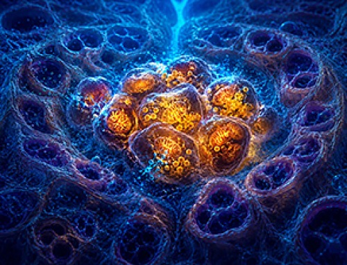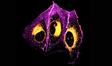
Lipids are difficult to detect with light microscopy. Using a new chemical labeling strategy, a Dresden-based team led by André Nadler at the Max Planck Institute of Molecular Cell Biology and Genetics (MPI-CBG) and Alf Honigmann at the Biotechnology Center (BIOTEC) of TU Dresden has overcome this limitation, enabling new insights into lipids.
The researchers were able to answer a long-standing question: how do cells transport specific lipids to the membranes of their target organelles? The new lipid-imaging technique will help understand the role of lipid transport in health and disease. The findings were published in the journal Nature.
Lipid molecules, or fats, are crucial to all forms of life. Cells need lipids to build membranes, separate and organize biochemical reactions, store energy, and transmit information.
Every cell can create thousands of different lipids, and when they are out of balance, metabolic and neurodegenerative diseases can arise. It is still not well understood how cells sort different types of lipids between cell organelles to maintain the composition of each membrane.
A major reason is that lipids are difficult to study, since microscopy techniques to precisely trace their location inside cells have so far been missing.
In a long-standing collaboration, Nadler, a chemical biologist at the Max Planck Institute of Molecular Cell Biology and Genetics (MPI-CBG) in Dresden, Germany, teamed up with Honigmann, a bioimaging specialist at Biotechnology Center (BIOTEC) at the TU-Dresden University of Technology, to develop a method that enables visualizing lipids in cells using standard fluorescence microscopy.
After the first successful proof of concept, the duo brought mass-spectrometry expert Andrej Shevchenko (MPI-CBG), Björn Drobot at the Helmholtz-Zentrum Dresden-Rossendorf (HZDR), and the group of Martin Hof from the J. Heyrovsky Institute of Physical Chemistry in Prague on board to study how lipids are transported between cellular organelles.
Artificial lipids under the sunbed
“We started our project with synthesizing a set of minimally modified lipids that represent the main lipids present in organelle membranes. These modified lipids are essentially the same as their native counterparts, with just a few different atoms that allowed us to track them under the microscope,” explains Kristin Böhlig, a Ph.D. student in the Nadler group and chemist who was in charge of creating the modified lipids.
The modified lipids mimic natural lipids and are “bifunctional,” which means they can be activated by UV light, causing the lipid to bind or crosslink with nearby proteins. The modified lipids were loaded in the membrane of living cells and, over time, transported into the membranes of organelles. The researchers worked with human cells in cell culture, such as bone or intestinal cells, as they are ideal for imaging.
“After the treatment with UV light, we were able to monitor the lipids with fluorescence microscopy and capture their location over time. This gave us a comprehensive picture of lipid exchange between cell membrane and organelle membranes,” concludes Kristin.
In order to understand the microscopy data, the team needed a custom image analysis pipeline. “To address our specific needs, I developed an image analysis pipeline with automated image segmentation assisted by artificial intelligence to quantify the lipid flow through the cellular organelle system,” says Juan Iglesias-Artola, who did the image analysis.
Speedy lipid transport by proteins
By combining the image analysis with mathematical modeling done by Drobot at the HZDR, the research team discovered that between 85% and 95% of the lipid transport between the membranes of cell organelles is organized by carrier proteins that move the lipids, rather than by vesicles.
This non-vesicular transport is much more specific with regard to individual lipid species and their sorting to the different organelles in the cell. The researchers also found that the lipid transport by proteins is ten times faster than by vesicles. These results imply that the lipid compositions of organelle membranes are primarily maintained through fast, species-specific, non-vesicular lipid transport.
In a parallel set of experiments, the group of Shevchenko at the MPI-CBG used ultra-high-resolution mass spectrometry to see how the different lipids change their structure during the transport from the cell membrane to the organelle membrane.
A boost for lipids in cell biology and disease
This new approach provides the first-ever quantitative map of how lipids move through the cell to different organelles. The results suggest that non-vesicular lipid transport has a key role in the maintenance of each organelle membrane composition.
Honigmann, research group leader at the BIOTEC, says, “Our lipid-imaging technique enables the mechanistic analysis of lipid transport and function directly in cells, which has been impossible before. We think that our work opens the door to a new era of studying the role of lipids within the cell.”
Imaging of lipids will allow further discoveries and help to reveal the underlying mechanisms in diseases caused by lipid imbalances. The new technique could potentially help to develop new druggable targets and therapeutic approaches for lipid-associated diseases, such as nonalcoholic fatty liver disease.
‘We knew that we were onto something big’
Nadler, research group leader at MPI-CBG, looks back at the start of the study: “Imaging lipids in cells has always been one of the most challenging aspects of microscopy. Our project was no different. Alf Honigmann and I started discussing about solving the lipid imaging problem as soon as we got hired in close succession at MPI-CBG in 2014/15 and we quickly decided to go for it.
“It still took us almost five years from the start of the project to the point in autumn 2019 when the two of us finally produced a sample with a beautiful plasma membrane stain. That’s when we knew that we were onto something big. As a reward, certain well-known global events meant we were required to shut down our laboratories a few months later.
“In the end, the delay was for the best. Before the revolution in the use of artificial intelligence in image segmentation, we would not have been able to properly quantify the imaging data, so our conclusions would have been much more limited.”
Researchers still need to determine which lipid-transfer proteins drive the selective transport of different lipid species. They also need to identify the energy sources that power lipid transport and ensure that each organelle keeps its own unique membrane composition.
More information: Juan M. Iglesias-Artola et al, Quantitative imaging of lipid transport in mammalian cells, Nature (2025). DOI: 10.1038/s41586-025-09432-x
Journal information: Nature

News
Cutting Two Amino Acids Slowed Prostate Cancer in Mice
A newly identified link between amino acid metabolism and cholesterol production may help prostate tumors adapt to hormone therapy. Prostate cancer can find ways around treatments designed to deprive tumors of the hormones they [...]
Largest-Ever Physics Survey Raises New Doubts About Our Model of the Universe
Physicists around the world remain deeply divided on key mysteries of the universe, from dark matter to quantum gravity. The standard cosmological model failed to gain majority support, and no leading theory dominated the [...]
Scientists Just Overturned a 100-Year-Old Belief About Bacteria in the Lungs
New findings raise questions about the role of microbes living in the lungs. More than 35 trillion bacteria live throughout the human body, forming microbiomes in the gut, mouth, lungs, skin, and urogenital tract. [...]

GHCE Concept
From the preface of the book Global Health Care Equivalency in the Age of Nanotechnology, Nanomedicine and Artificial Intelligence, Edited by Frank Boehm: Since the publication of my first book (Nanomedical Device and Systems [...]
Novartis, Ionis drug failure spurs questions
Pelacarsen didn’t protect heart health despite lowering levels of a protein particle, “Lp(a),” in a large clinical trial — a result with important implications for cardiovascular drug research. Dive Brief: An RNA drug from [...]
New injectable treatment helps the brain rebuild after stroke
Biomedical engineers at Duke University have created an injectable biomaterial that may help the brain recover from damage left behind by an ischemic stroke. In experiments with mice, the material transformed the cavity created [...]
Scientists Discover a Hidden “Immune Organ” Inside the Skull
Researchers discovered lymph node-like immune hubs inside skull bone marrow that appear to act as rapid-response centers for the brain. For decades, the brain was thought to operate largely apart from the immune system. [...]
Engineered tRNAs and lipid nanoparticles target nonsense mutation cystic fibrosis
Researchers have developed a potential new approach for treating a form of cystic fibrosis caused by so-called nonsense mutations, combining chemically modified transfer RNAs with lipid nanoparticles designed to deliver the therapy directly to [...]
New pancreatic cancer drug carries a $39,800 monthly list price
A groundbreaking treatment for one of the most common forms of pancreatic cancer has been approved in pill form by the FDA. Revolution Medicines’ oral tablet daraxonrasib, branded as Rasonque, reduced the risk of [...]
Researchers Have Discovered a New Way To Reduce Chronic Nerve Pain
A cancer-linked protein called BRAF may help drive chronic nerve pain, and existing cancer drugs targeting it reduced pain sensitivity in preclinical models. Chronic nerve pain can persist long after an injury and often [...]
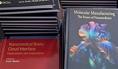
Our books now available worldwide!
Online Sellers other than Amazon, Routledge, and IOPP Indigo Global Health Care Equivalency in the Age of Nanotechnology, Nanomedicine and Artifcial Intelligence Global Health Care Equivalency In The Age Of Nanotechnology, Nanomedicine And Artificial [...]

Quantum-Enabled Regenerative Health: Reimagining Wellness, Precision Health and Longevity Medicine
Introduction Healthcare is approaching a frontier where the quantum portfolio could influence not only how disease is diagnosed and treated, but how health itself is measured, modeled, predicted and preserved. Quantum computing, quantum simulation, [...]

FDA Clears First-of-Its-Kind Nonmedication Treatment for PTSD
The FDA has cleared a system that uses brain activity data to personalize magnetic stimulation for PTSD, adding a new nonmedication treatment option. Every day in the United States, approximately 17.5 veterans die by suicide, [...]
FDA approves breakthrough drug to treat advanced pancreatic cancer
The Food and Drug Administration (FDA) approved on Wednesday a drug that could extend the survival of those with metastatic pancreatic cancer. The drug, called daraxonrasib, will be sold under the brand name Rasonque [...]
AI Decodes a Hidden DNA Signal Linked to Disease-Causing Mutations
Machine learning identifies the likely “initiator” and enables new predictions about DNA mutations that can cause disease. Every human cell depends on tens of thousands of genes being switched on at the right time [...]

Pope Leo Urges Global Response to Congo’s Deadliest Ebola Outbreak
Pope Leo called for international action to address the Ebola outbreak in the Democratic Republic of Congo. The epidemic has claimed over 2,500 lives and is the nation's largest recorded outbreak. The Pope emphasized [...]