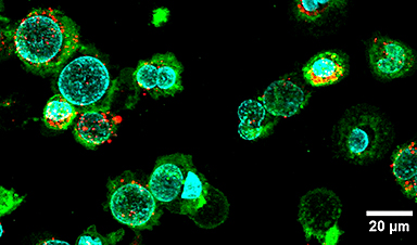
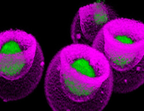
| Particles known as extracellular vesicles play a vital role in communication between cells and in many cell functions. Released by cells into their environment, these “membrane particles” consist of a cellular membrane carrying a cargo of specific signaling molecules, proteins, nucleic acids and lipids. Unfortunately, only tiny quantities of the vesicles are formed spontaneously by cells. | |
Extracellular vesicles for medical applications |
|
| The contents of these extracellular vesicles vary depending on the origin and condition of the cell, as do the proteins that are anchored to the vesicle surface. Researchers use these properties to develop new techniques for diagnosing cancer, for example, based on the analysis of extracellular vesicles isolated from blood samples. | |
| Extracellular vesicles could also play a key role in the development of next-generation therapeutics. As the vesicles are of natural origin, they are biocompatible and can trigger a wide range of different reactions in the body. | |
| Researchers therefore hope to use the particles to influence the immune system – for example, in order to destroy cancer cells. Until now, however, one major challenge has been the reproducible production of the large quantities of homogeneous vesicles needed for such studies. |
A faster route to more particles |
|
| Now, a team of researchers led by Professor Jörg Huwyler from the Department of Pharmaceutical Sciences and the Swiss Nanoscience Institute (SNI) of the University of Basel has developed a highly efficient preparation method for extracellular vesicles that delivers up to 100 times more particles per cell and hour than conventional methods. They describe the new method in the journal Communications Biology (“High efficiency preparation of monodisperse plasma membrane derived extracellular vesicles for therapeutic applications”). | |
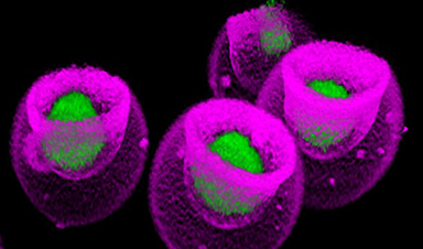
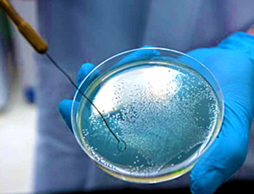
| “We start the preparation process by cultivating cancer cells, in which we induce cell death by adding chemical stressors,” explains Claudio Alter, first author of the study and a doctoral student at the SNI PhD School. “The cells then form vesicles, which detach from the parent cell after a few hours.” | |
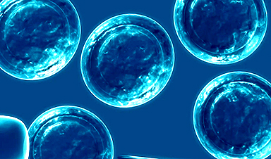
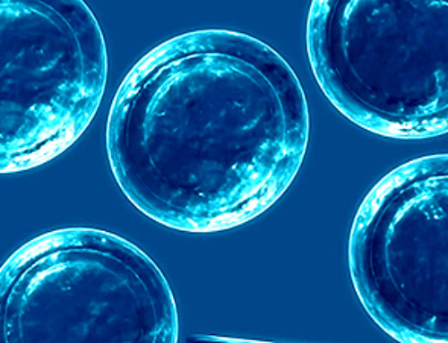
| With a diameter of 1 to 3 micrometers, these giant plasma membrane vesicles are far too big for therapeutic applications. In the newly developed process, they are therefore pressed through a filter membrane multiple times in order to reduce their size. “After multiple filter passes, we obtain a homogeneous solution of nano plasma membrane vesicles (nPMV) with a diameter of 120 nanometers – precisely what we need for subsequent applications,” explains Alter. | |
Different origin, different applications |
|
| The team of researchers then characterized these nPMVs and compared their size, homogeneity, and protein and lipid cargo with those of exosomes – currently the most commonly used extracellular vesicles. They also investigated how well the nPMVs interact with other cells. In these analyses, the nano plasma membrane vesicles showed similar properties to exosomes. | |
| “Their specific cargo and the presence of membrane-bound markers derived from the parent cell line offers the possibility to use nPMVs for therapeutic purposes,” says Jörg Huwyler. “At present, we’re primarily thinking of a stimulation of the immune system – for example, in vaccination or in immunotherapy treatments for cancer.” |
News
How Are Hydrogels Shaping the Future of Biomedicine?
Hydrogels have gained widespread recognition and utilization in biomedical engineering, with their applications dating back to the 1960s when they were first used in contact lens production. Hydrogels are distinguished from other biomaterials in [...]
Nanovials method for immune cell screening uncovers receptors that target prostate cancer
A recent UCLA study demonstrates a new process for screening T cells, part of the body's natural defenses, for characteristics vital to the success of cell-based treatments. The method filters T cells based on [...]
New Research Reveals That Your Sense of Smell May Be Smarter Than You Think
A new study published in the Journal of Neuroscience indicates that the sense of smell is significantly influenced by cues from other senses, whereas the senses of sight and hearing are much less affected. A popular [...]
Deadly bacteria show thirst for human blood: the phenomenon of bacterial vampirism
Some of the world's deadliest bacteria seek out and feed on human blood, a newly-discovered phenomenon researchers are calling "bacterial vampirism." A team led by Washington State University researchers has found the bacteria are [...]
Organ Architects: The Remarkable Cells Shaping Our Development
Finding your way through the winding streets of certain cities can be a real challenge without a map. To orient ourselves, we rely on a variety of information, including digital maps on our phones, [...]
Novel hydrogel removes microplastics from water
Microplastics pose a great threat to human health. These tiny plastic debris can enter our bodies through the water we drink and increase the risk of illnesses. They are also an environmental hazard; found [...]
Researchers Discover New Origin of Deep Brain Waves
Understanding hippocampal activity could improve sleep and cognition therapies. Researchers from the University of California, Irvine’s biomedical engineering department have discovered a new origin for two essential brain waves—slow waves and sleep spindles—that are critical for [...]
The Lifelong Cost of Surviving COVID: Scientists Uncover Long-Term Effects
Many of the individuals released to long-term acute care facilities suffered from conditions that lasted for over a year. Researchers at UC San Francisco studied COVID-19 patients in the United States who survived some of the longest and [...]
Previously Unknown Rogue Immune Key to Chronic Viral Infections Discovered
Scientists discovered a previously unidentified rogue immune cell linked to poor antibody responses in chronic viral infections. Australian researchers have discovered a previously unknown rogue immune cell that can cause poor antibody responses in [...]
Nature’s Betrayal: Unmasking Lead Lurking in Herbal Medicine
A case of lead poisoning due to Ayurvedic medicine use demonstrates the importance of patient history in diagnosis and the need for public health collaboration to prevent similar risks. An article in CMAJ (Canadian Medical Association [...]
Frozen in Time: How a DNA Anomaly Misled Scientists for Centuries
An enormous meteor spelled doom for most dinosaurs 65 million years ago. But not all. In the aftermath of the extinction event, birds — technically dinosaurs themselves — flourished. Scientists have spent centuries trying [...]
‘Mini kidneys’ reveal new insights into metabolic defects in polycystic kidney disease
Scientists at Nanyang Technological University, Singapore (NTU Singapore) have successfully grown 'mini kidneys' in the lab and grafted them into live mice, revealing new insights into the metabolic defects and a potential therapy for [...]
Decoding the Origin of Life: Scientists Solve Early Earth RNA Puzzle
Recent research illustrates how RNA molecules’ chemical characteristics might have played a crucial role in the development of complex life forms. How did complex life manage to evolve on the early, inhospitable Earth? Initially, [...]
Improving infectious disease testing with gold nanoparticles
By harnessing the power of composite polymer particles adorned with gold nanoparticles, a group of researchers have delivered a more accurate means of testing for infectious diseases. Details of their research was published in the [...]
New micromaterial releases nanoparticles that selectively destroy cancer cells
Researchers have developed micromaterials made up only of proteins, capable of delivering over an extended period of time nanoparticles that attack specific cancer cells and destroy them. The micromaterials mimic natural secretory granules found [...]
Alzheimer’s Breakthrough: Scientists Make Revolutionary Leap
Dementia is a major health issue worldwide in the 21st century, impacting over 50 million people globally. This figure is expected to soar to 152 million by 2050, as the global population ages. Alzheimer’s disease (AD) [...]
