In an article recently published in the open-access MDPI journal nanomaterials, researchers investigated the mechanism of interaction between nanoscale materials and specific cells for biomedical applications, primarily related to targeted delivery and tracing.
Here, the localization, cellular uptake quantity, and response time of gold nanorods in MCF-7 and SKBR-3 breast cancer cells were measured.
Background
The amalgamation of biotechnology and nanotechnology has been a quite fascinating field of research for the last few years.
Currently, nanomaterials are extensively used in biomedical applications such as drug delivery, magnetic or fluorescent bio-imaging, tissue engineering, tracing pathogens in sensitive organs, cancer chemotherapy, and many more. However, each nanomaterial interacts differently with a particular type of cell.
Many studies have shown cellular uptake of gold nanorods by cancer cells. MCF-7 and SKBR-3 cancer cell lines represent HER2-negative and HER2-positive breast cancer models, respectively. These cells intake gold nanorods through a mechanism occurring at the surface of the cell membrane, called endocytosis.
Inductively coupled plasma mass spectrometry (ICP-MS) can measure the efficiency of targeted delivery and cellular uptake of these nanoparticles, while transmission electron microscopy (TEM) can be used to investigate their localization.
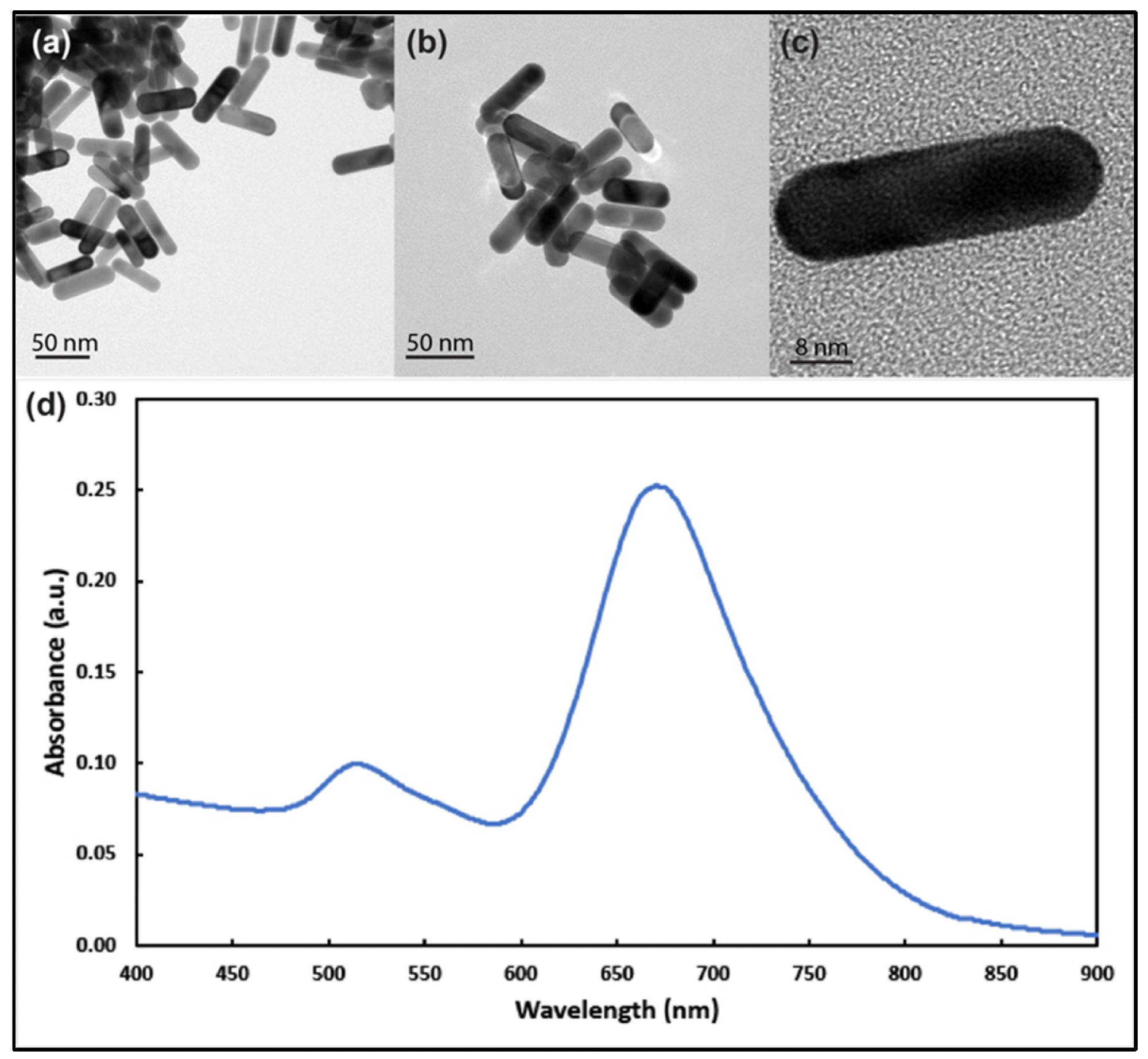
Figure 1. Transmission electron microscope images of gold nanorods with aspect ratio around 3 at different magnifications (a–c). (d) UV–visible spectrum analysis of the prepared gold nanorods showing longitudinal and transverse plasmons around 690 nm and 514 nm, respectively. © White, B., White, M., Nima Alsudani, Z., Watanabe, F., Biris, A., Ali, N., (2022)
About the Study
In this article, researchers investigated the interaction mechanism of gold nanorods (Au NRs) in two types of breast cancer cell lines namely, MCF-7 and SKBR-3. More than 200 Au NRs with an average length and diameter of 36 and 12 nm, respectively, were synthesized using the silver ion-assisted seed-mediated method. Subsequently, MCF-7 and SKBR-3 cells were cultured in a conducive medium and allowed to bind with different concentrations of Au NRs for 1 and 2 days.
The excess Au NRs were washed away from the treated cancels using phosphate buffer saline (PBS) solution followed by treating with the (3-(4,5-dimethylthiazol-2-yl)-2,5-diphenyltetrazolium bromide) (MTT) reagent. Finally, ICP-MS and TEM analyses were performed to investigate the interaction mechanism.
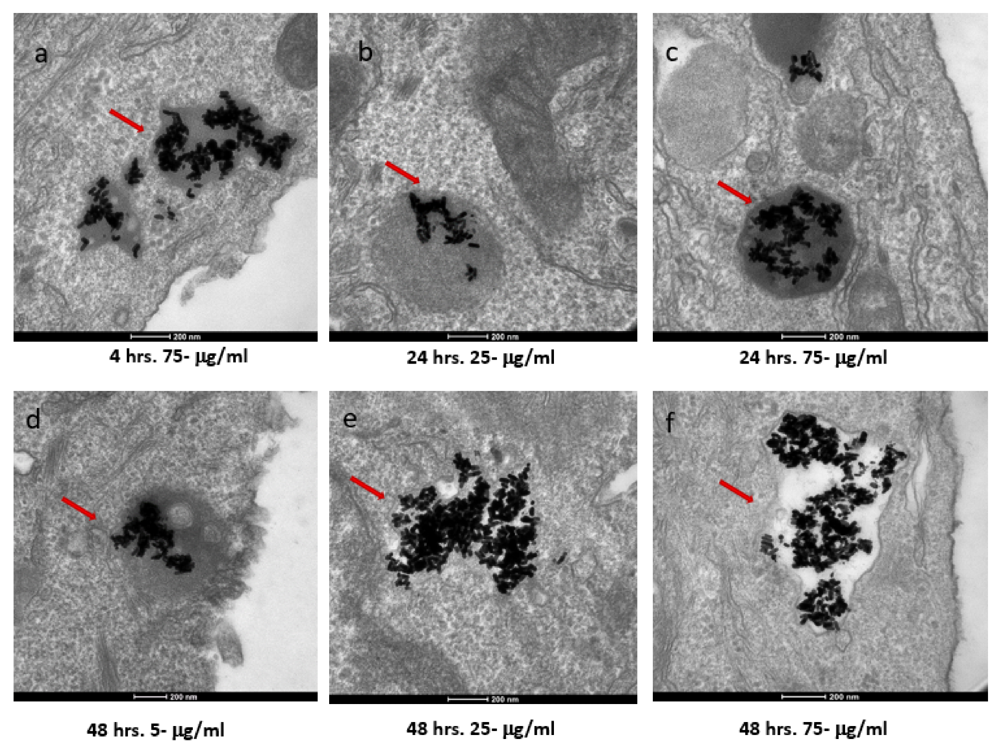
Figure 2. TEM images of AuNRs in SKBR-3 cells at different time points and concentrations. SKBR-3 cells were treated with 5, 25, and 75 μg/mL of AuNRs for 1, 4, 24, and 48 h. Internalization of nanorods was observed starting at 4hourincubation with 75 μg/mL (a). After 24hourof incubation, internalization was observed with the 25 μg/mL (b) and 75 μg/mL concentrations (c). For cells incubated for 48 h, we observed internalization with 5, 25, and 75 μg/mL concentrations (d, e, and f, respectively). Red arrows point to gold nanorods localized in SKBR-3 cells. Electron photomicrographs shown are representative of at least three independent experiments with similar conditions. © White, B., White, M., Nima Alsudani, Z., Watanabe, F., Biris, A., Ali, N., (2022)
Observations
TEM images showed Au NRs had longitudinal and transverse plasmon resonances at around 680 and 514 nm, respectively. The average zeta potential of the NRs was −47.18 ± 4.26 mV. Au NRs did not show any cytotoxicity in healthy human cells. MTT methods indicated that the MCF-7 cells were less sensitive to Au NR exposure than the SKBR-3 cells for longer exposure times at higher concentrations. The SKBR-3 cells exhibited a drop in cell vitality due to higher sensitivity, whereas the MCF-7 cells had almost no drop in cell viability.
Both types of cells demonstrated a different pattern in cellular uptake of Au NRs for different exposure times and concentrations. The SKBR-3 cells had a constant cellular uptake between one to four hours, but 25 μg/mL Au NRs concentration had a higher uptake than the 75 μg/mL sample, i.e. lower concentration had a higher cellular uptake. In contrast, in MCF-7 cells, the 75 μg/mL AuNR-treated cell samples exhibited higher cellular uptake than 5 and 25 μg/mL samples, i.e. higher concentration samples had higher cellular uptake than lower concentration samples.
Furthermore, TEM analysis showed that the Au NRs interacted with the cell membrane of both cancer cells starting from one to 48 hours of exposure time. However, the Au NRs entered into the cell (i.e. internalized) for the samples with concentrations of 25 and 75 μg/mL. The 5 μg/mL concentration samples were never internalized. Moreover, the minimum exposure time for internalization was one hour and four hours for the MCF-7 and SKBR-3 cells, respectively.
The mechanism for the cellular uptake of the Au NRs was primarily micropinocytosis instead of receptor-mediated endocytosis. These Au NRs-carrying macropinosomes further interacted with the lysosomes inside the cells.
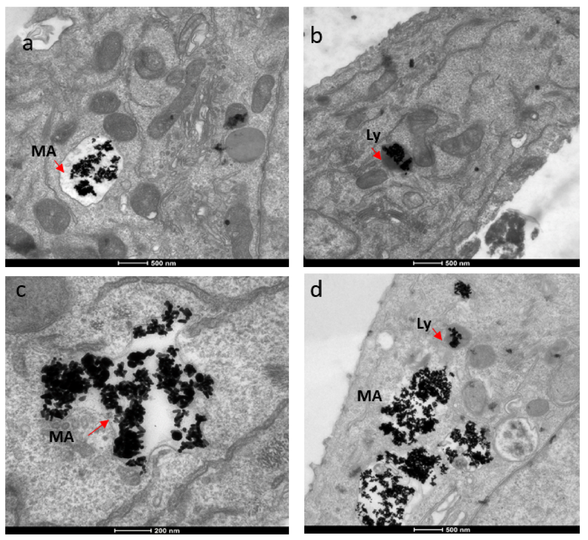
Figure 3. TEM images showing localization of gold nanorods in macropinosomes and lysosomes in both SKBR-3 and MCF-7 cell lines. AuNR localization in (a) macropinosomes (MA) and (b) lysosomes (Ly) of SKBR-3 cells. (c) Macropinosomes (MA) containing gold nanorods in MCF-7 and (d) lysosomes (Ly) containing gold nanorods in MCF-7. Red arrows point out macropinosomes and lysosomes containing gold nanorods in each image. Electron photomicrographs shown are representative of at least three independent experiments with similar conditions. © White, B., White, M., Nima Alsudani, Z., Watanabe, F., Biris, A., Ali, N., (2022)
Conclusions
To conclude, the researchers of this study investigated the interaction of Au NRs in two types of breast cancer cell lines, i.e. MCF-7 and SKBR-3. The shape, size, and zeta potential of Au NRs had different effects on cell vitality and toxicity of different cancer cells. Additionally, both cell types had different patterns of cellular uptake for Au NRs at different concentrations and exposure times.
The Au NRs did not enter into the cancer cells for samples with lower concentrations of NRs or lower exposure time. Also, the cellular uptake mechanism was primarily driven by micropinocytosis. These findings will help improve biomedical applications related to targeted drug delivery, tracing, and bio-imaging involving nanomaterials.
News

Our books now available worldwide!
Online Sellers other than Amazon, Routledge, and IOPP Indigo Global Health Care Equivalency in the Age of Nanotechnology, Nanomedicine and Artifcial Intelligence Global Health Care Equivalency In The Age Of Nanotechnology, Nanomedicine And Artificial [...]

Younger Generations Are Aging Faster – and It May Be Fueling a Surge in Cancer
Younger generations may be aging biologically faster than those before them, and that shift could help explain rising rates of cancer at younger ages. For decades, cancer was viewed largely as a disease of [...]

Using Cannabis Could Raise Your Stroke Risk by 37%, Massive Study Reveals
Large-scale evidence suggests cannabis, cocaine, and amphetamines may directly raise stroke risk, including in younger adults. As recreational drug use becomes increasingly common, researchers are uncovering evidence that its health consequences may extend far beyond [...]

Could Vitamin C Be the Secret to Keeping Your Brain Younger?
Lower vitamin C levels were linked to reduced brain volume and weaker neural connectivity in older adults, suggesting a potential connection between nutrition and brain health. Could a common vitamin help preserve the brain [...]

This Deadly Disease Was Wiping Out Humans 5,500 Years Ago
A new study suggests plague was already a deadly threat 5,500 years ago, striking small hunter-gatherer communities long before cities and agriculture emerged. For centuries, plague has been remembered as the disease that devastated [...]

China closing in but US leads in biotech quality, commercial reach, survey finds
SAN DIEGO, June 22 (Reuters) - China, which now conducts more clinical drug trials, opens new tab than the U.S., still lags in the quality and commercial reach of its biomedical science, according to a recent survey, opens new [...]

New method generates renewable supply of progenitor immune cells
In a paper published in Cell, a USC Stem Cell-led team reports a new way of generating a renewable and expandable supply of the progenitor cells that give rise to macrophages. These immune cells help [...]

Scientists Just Discovered a Cellular Survival System That Was Never Supposed To Exist
A surprising backup pathway allows cells to make a crucial amino acid when their primary machinery fails. For decades, biologists believed cells had only one way to access a molecule they cannot live without. New [...]

Artificial cells gain porous membranes, enabling lab reactions and drug release
Artificial cells created in the laboratory offer a wide range of potential applications. Until now, however, their membranes—unlike those of real cells—have been virtually impermeable. Researchers at the Max Planck Institute for Polymer Research, [...]

Popular Weight-Loss Drugs Like Ozempic Linked to Lower Breast Cancer Risk
Ozempic and similar weight-loss drugs were linked to a striking 30% reduction in breast cancer risk in a study of more than 110,000 women. Popular weight-loss and diabetes medications such as Ozempic, Wegovy, Mounjaro, [...]

Stanford Scientists Discover Explosive New Type of Immune Cell
Scientists studying the remarkable regenerative abilities of planarian flatworms have uncovered a previously unknown type of immune cell with an unusually destructive defense strategy. What if an immune cell could wipe out nearby threats [...]

Big Pharma-backed SonoThera sounds off with $125M series B for bubble-based genetic delivery
Bay Area biotech SonoThera is bubbling to a clinical boil after raising a $125 million series B with the backing of some of the biggest names in pharma. Vida Ventures led the raise, with the venture [...]

Joint initiative of 5 EU countries calls for ‘unified approach’ to pharma framework amid US drug pricing pressure
With drug pricing pressure building from the U.S., a healthcare-focused consortium of five European countries is calling for a “unified approach” to strengthen Europe’s pharmaceutical framework and access to innovative medicines. Belgium, the Netherlands, [...]

Molecular Manufacturing: The Future of Nanomedicine – New book from NanoappsMedical Inc.
This book explores the revolutionary potential of atomically precise manufacturing technologies to transform global healthcare, as well as practically every other sector across society. This forward-thinking volume examines how envisaged Factory@Home systems might enable the cost-effective [...]

NanoMedical Brain/Cloud Interface – Explorations and Implications. A new book from Frank Boehm
New book from Frank Boehm, NanoappsMedical Inc Founder: This book explores the future hypothetical possibility that the cerebral cortex of the human brain might be seamlessly, safely, and securely connected with the Cloud via [...]

New book from Nanoappsmedical Inc. – Global Health Care Equivalency
A new book by Frank Boehm, NanoappsMedical Inc. Founder. This groundbreaking volume explores the vision of a Global Health Care Equivalency (GHCE) system powered by artificial intelligence and quantum computing technologies, operating on secure [...]