A group of researchers recently published a paper in the journal Materials that demonstrated the viability of using vancomycin-functionalized gold nanoparticles (V-GNPs) against pathogenic bacterial strains.
Background
Antibiotic resistance among pathogenic bacteria is rising at a remarkable rate, which poses a significant risk to human health. For instance, vancomycin, a glycopeptide antibiotic, is typically used for treating bacterial infections such as methicillin-resistant Staphylococcus aureus (MRSA). However, the excessive use of vancomycin has led to the emergence of vancomycin-resistant bacterial strains.
Innovative strategies such as the use of nanoparticles (NPs) hold the potential to combat antibiotic-resistant bacterial infections.
The characteristics of NPs, such as surface chemistry, size, and shape, can be manipulated easily, which makes them suitable for fighting bacterial infections.
Metallic NPs, such as GNPs, are specifically being evaluated in different studies for their potential in treating bacterial infections.
Several studies have shown that the antibacterial activities of antibiotics can be enhanced by conjugating GNPs with antibiotics.
In this study, researchers investigated the ability of GNPs to enhance the antibacterial efficacy of the antibiotic vancomycin against different bacterial strains.
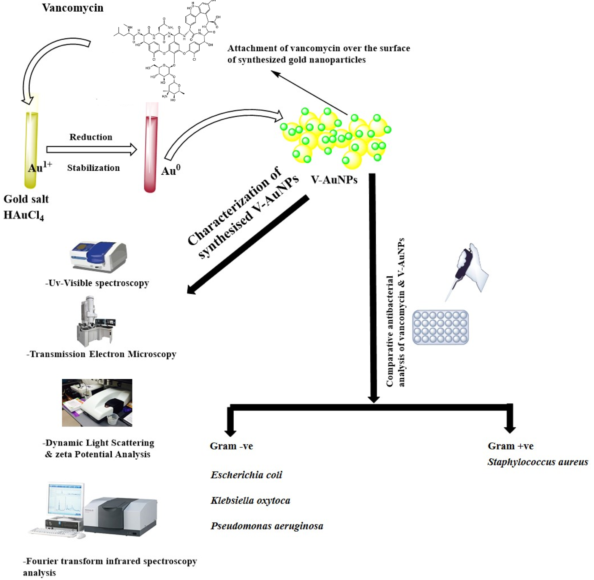
Figure 1. Schematic representation of vancomycin-mediated synthesis of gold nanoparticles (V-GNPs), their characterization, and antibacterial testing. © Hagbani, T.A., Yadav, H., Moin, A. et al. (2022)
The Study
Gold (III) chloride trihydrate (HAuCl4·3H2O), phosphate buffer, and vancomycin were used for preparing V-GNPs in the study.
In the preparation, 1, 0.75, 0.50, or 0.25 mg/mL of vancomycin was mixed with a 3 mL reaction mixture containing 50 mM phosphate buffer with a pH value of 7.4 and 1 mM HAuCl4·3H2O, and the resultant mixture was incubated at 60, 50, 40, and 30 oC, respectively, for 48 h.
The synthesized V-GNPs were obtained by centrifugation of the reaction mixture for 30 min at 30,000× g and the collected NPs were treated with 50% v/v ethanol in order to eliminate unattached materials.
The Gram-positive bacterial strain of Staphylococcus aureus and the Gram-negative strains of Pseudomonas aeruginosa, Klebsiella oxytoca, and Escherichia coli were used in the study to evaluate the antibacterial efficacy of V-GNPs.
Each bacterial strain was maintained and cultivated in Mueller Hinton (MH) agar media at 37 oC.
A Shimadzu UV-1601 spectrophotometer was employed to obtain the ultraviolet ultraviolet-visible (UV-vis) spectra of the prepared V-GNPs in the range of 200-800 nm at 1 nm resolution.
The average particle size of the synthesized V-GNPs was determined using a dynamic light scattering particle (DLS) size analyzer, while the zeta-potential of the synthesized NPs was measured using a Malvern Zetasizer Nano-ZS.
A transmission electron microscopy (TEM) operating at an accelerating voltage of 80 kV was used to assess the homogeneity of the prepared V-GNPs, while Fourier transform infrared (FTIR) spectroscopy was performed to evaluate the conformational changes after vancomycin was loaded on the surface of GNPs.
The FTIR spectra were acquired using the potassium bromide (KBr) pellet method with a Shimadzu FTIR-8201 spectrometer within the range of 4000–400 cm-1 at a resolution of 4 cm-1.
The agar well diffusion methodology was utilized to evaluate the efficacy of prepared V-GNPs and pure vancomycin.
The in vitro antibacterial activity of pure vancomycin and V-GNPs was assessed using the minimal inhibitory concentration (MIC) method. The loading efficiency of V-GNPs was also calculated. All findings in the study were analyzed using GraphPad Prism through a one-way analysis of variance (ANOVA).
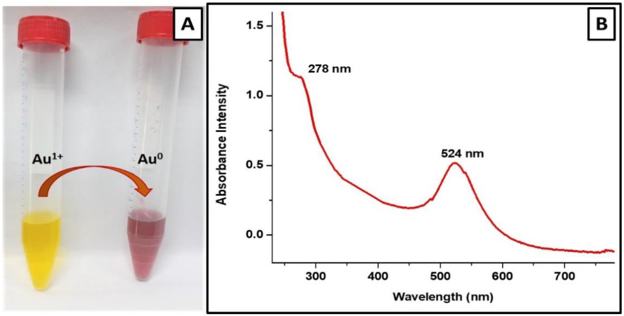
Figure 2. Characterization of V-GNPs: (A) color change from light yellow to ruby red resulted from SPR; (B) UV–Visible spectra (SPR band at 524 nm). © Hagbani, T.A., Yadav, H., Moin, A. et al. (2022)
Observations
GNPs with good physicochemical properties were synthesized at a pH of 7.4, a temperature of 40 oC, and a vancomycin concentration of 250 µg/mL.
The color of the gold salt solution changed from pale yellow to ruby red following the addition of 250 µg/mL vancomycin antibiotic, indicating the formation of V-GNPs.
The UV-Vis spectra of the prepared V-GNPs revealed the presence of a surface plasmon resonance peak at 524 nm.
The TEM micrographs showed that the synthesized V-GNPs were monodispersed, homogenous, and spherical in shape with an average size of 24 nm. No agglomeration was observed in the TEM micrographs, indicating the suitability of vancomycin as a stabilizing agent.
The hydrodynamic diameter of the V-GNPs was estimated as 77 nm by the DLS.
The zeta potential of the prepared V-GNPs was -18mV, indicating good stability of GNPs. Observations from the FTIR spectroscopy indicated that the vancomycin was efficiently loaded on the GNP surfaces, and the loading efficiency was 86.2%.
Both V-GNPs and pure vancomycin significantly inhibited bacterial growth. However, V-GNPs demonstrated more effective antibacterial activity compared to pure vancomycin even at a lower concentration of vancomycin.
The in vitro antibacterial studies demonstrated that the antibacterial activities of the synthesized V-GNPs were 1.6-, 1.8-, 1.6-, and 1,4-fold higher than pure vancomycin against Staphylococcus aureus, Pseudomonas aeruginosa, Klebsiella oxytoca, and Escherichia coli, respectively.
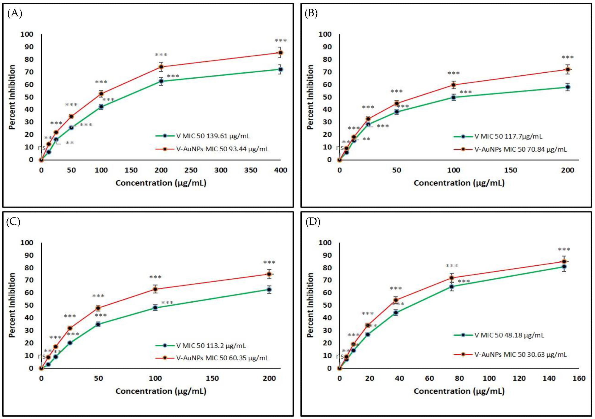
Figure 3. Determination of the minimum inhibitory concentration (MIC) of vancomycin (V) and V-GNPs against (A) Escherichia coli; (B) Klebsiella oxytoca; (C) Pseudomonas aeruginosa; (D) Staphylococcus aureus. The experiment was repeated in triplicate, and the data shown are the means ± standard errors. Significantly different from control at ** p < 0.01, *** p < 0.001; non-significant from the control at ns p > 0.05. © Hagbani, T.A., Yadav, H., Moin, A. et al. (2022)
Significance of the Study
Taken together, the findings of the study demonstrated that V-GNPs represent a more suitable alternative compared to pure vancomycin against pathogenic bacterial strains.
However, in vivo investigations must be performed to assess the toxicity of V-GNPs before the synthesized vancomycin nanoformulations can be used to treat infections. Nevertheless, this study can act as a starting point for developing better antibacterial nanoformulations.
News

Electrostatic Discharge Boosts Triboelectric Nanogenerator Current and Enables DC Output
Controlled electrical discharges could enable triboelectric nanogenerators to achieve higher peak currents, extending nano-enabled energy harvesting into chemical processing and self-powered sensing. Paper: Electrostatic discharge as a breakthrough strategy for triboelectric nanogenerators. A new review [...]
Swiss laboratory uses old drugs against rare diseases
Researchers at the University of Geneva are combing through collections of approved drugs to find new therapies for rare diseases – with some success. This approach is gaining traction around the world, while pharmaceutical [...]

Nanozyme Aptasensors Show Promise for Faster Food, Health, and Environmental Testing
By pairing robust artificial enzymes with highly selective aptamers, nanozyme aptasensors could help detect disease biomarkers, pathogens, and contaminants faster, but the review shows that real-world deployment still depends on overcoming matrix interference, biofouling, [...]

Paralyzed Man Feels Sensation Again With Brain Stimulation Device
Aneuroprosthetic system has allowed a man with paralysis to grasp and lift objects and feel touch again. The device helped 42-year-old Keith Thomas of Massapequa, New York, who was paralyzed from the chest down [...]
Global Cancer Cases Could Surge 67% by 2050, New Report Warns
New data reveal major geographic disparities and highlight the urgent need for global action on prevention, early detection, and equitable access to treatment. For roughly one in five people worldwide, cancer will become part [...]

A Deadly Ebola-Like Virus Is Spreading. Are We Ready?
BU virologist Nancy Sullivan says the Bundibugyo outbreak in the Democratic Republic of the Congo underscores the need for broader outbreak preparedness. The death of a nurse marked the moment health officials recognized that [...]

Why Most Animal Viruses Never Become Human Pandemics
From receptor mismatch to risky human-animal interfaces, this article explains why spillover is common but true pandemic emergence remains rare. Introduction Humans are constantly exposed to animal viruses through farming, wildlife contact, and the [...]

Stem cell organoids repair heart microvessels in coronary artery disease models
A Stanford University team has shown that vascular organoids derived from human stem cells can repair the heart’s microvessel network in pigs with ischaemic heart disease – a proof-of-concept advancement that could open new therapeutic [...]

Goodbye GP waiting rooms, hello prevention at home
Prevention is suddenly everywhere in NHS reform. The recent £340m community pharmacy deal is moving more services onto the high street. Community Diagnostic Centres are being expanded, and the Neighbourhood Health Framework sets out [...]

Ebola control is weakened by mistrust and cultural insensitivity
Effective response depends on cooperation with communities and frontline workers, writes Zaeem ul Haq The current Bundibugyo Ebola outbreak in the Democratic Republic of the Congo (DRC) and Uganda is exposing dangerous gaps in [...]

Building the Brain Requires Millions of Dangerous DNA Breaks
Scientists discovered that building a healthy brain involves an unexpected step: young neurons routinely break and rapidly repair their own DNA. As the brain develops, newly formed nerve cells must travel through tightly packed tissue [...]

One Tiny Change May Explain How Viruses Jump From Bats to Humans
Scientists found that one tiny genetic change may determine whether a bat virus stays in bats or becomes a human threat. Most infectious disease outbreaks begin when a virus or other pathogen crosses from animals into [...]

Scientists Discover 250+ Genes That Could Lead to New Ways To Prevent Melanoma
The world’s largest study of mole genetics identified hundreds of genes tied to melanoma risk, uncovering potential new drug targets and paving the way for more accurate melanoma screening and prevention. Researchers at QIMR [...]
Breakthrough Diabetes Treatment Reprograms the Immune System
An engineered stem cell therapy reversed new-onset Type 1 diabetes in mice by shifting the immune system away from attacking insulin-producing cells. For more than a century, people with Type 1 diabetes have relied [...]
Taking the world’s temperature: WHO chief spotlights global health emergencies
Taking the world’s temperature on pressing health matters, WHO Director-General Tedros Adhanom Ghebreyesus provided the latest on current global challenges - and successes when it comes to international cooperation. “The outbreaks of hantavirus, Ebola and Marburg all show [...]
Scientists Create Tiny “Mini Livers” That Could One Day Replace Liver Transplants
Engineered tissue grafts could help perform key liver functions and benefit thousands of people living with liver failure. The liver is one of the body’s hardest-working organs, carrying out hundreds of vital jobs, from [...]