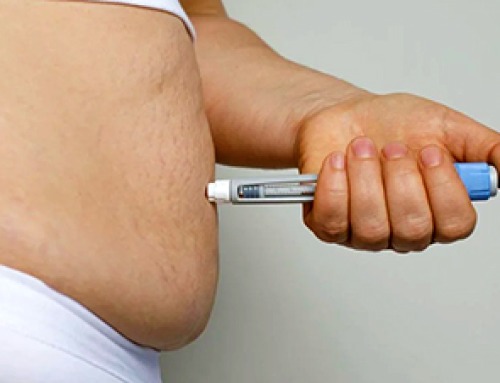
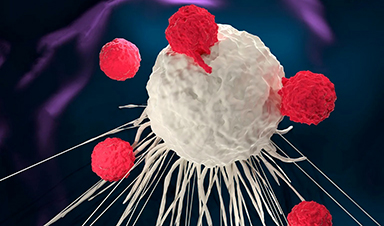
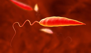
The blood-brain barrier blocks the entry of antibodies into the brain. This limits the potential use of antibody therapeutics to treat brain diseases, such as brain tumors.
In a new study published in the journal Frontiers in Cell and Developmental Biology, researchers at the University of Alabama at Birmingham report that the site-directed addition of an FDA-approved, biodegradable polymer at the hinge and near hinge regions of the therapeutic antibody trastuzumab effectively facilitated the brain delivery of this human monoclonal IgG1 antibody. Trastuzumab is used to treat breast cancer and several other cancers.
Preliminary work on this novel platform included in vitro and mouse-model experiments. Researchers say the delivery system still must be optimized and tested further, yet note their simple methodology converts antibody therapeutics to a brain-deliverable form that maintains the antibody’s medical functionality.
“The concerns of brain-entry haunt the development of brain-disease-targeting antibody therapeutics, impeding the medical translations of laboratory-generated antibodies to clinical practices,” said Masakazu Kamata, Ph.D., leader of the study and an associate professor in the UAB Department of Microbiology. “In this context, this simple methodology has great potential to serve as the platform to not only repurpose the current antibody therapeutics, but also encourage the design of novel antibodies, for the treatment of brain diseases.”
The biocompatible polymer used was poly 2-methacryloyloxyethyl phosphorylcholine, or PMPC, with chain lengths of 50, 100 or 200 monomers. The researchers had already discovered that this non-immunogenic polymer, which the FDA has approved as a coating material for transplantable devices, could bind to two receptors on brain microvascular endothelial cells composing the blood-brain barrier, and those cells could then move the polymer across the blood-brain barrier by transcytosis. Transcytosis is a specialized transport whereby extracellular cargo is brought inside the cell, shuttled across the cytoplasm to the other side of the cell, and then released.
The UAB researchers were able to cleave four interchain disulfide bonds in the trastuzumab IgG1 hinge and near hinge regions, creating thiol groups. Each thiol group was then conjugated to a chain of the PMPC to create trastuzumab molecules with one of the three chain lengths, which they denoted as Tmab-PMPC50, Tmab-PMPC100 and Tmab-PMPC200.
Each of these modified antibodies still maintained trastuzumab-specific binding to cells expressing the HER2 antigen, the target of trastuzumab. Both the Tmab-PMPC50 and the Tmab-PMPC100 were internalized into HER2-positive cells and promoted antibody-dependent cell death, which is the medical functionality by which trastuzumab kills HER2+ breast cancer cells.
The researchers then showed that PMPC conjugation of trastuzumab enhanced blood-brain barrier penetration through the epithelial cells on the blood-brain barrier via the transcytosis pathway. The translocatable Tmab-PMPC100 was the best at efficient blood-brain barrier penetration while retaining trastuzumab’s epitope recognition, the ability of the antibody to bind to its antigen target.
In a mouse model, both Tmab-PMPC100 and Tmab-PMPC200 were about fivefold better at brain penetration than native trastuzumab. In preliminary in vitro and mouse-model experiments, the polymer-modified trastuzumab did not induce neurotoxicity, did not show adverse effects in the liver, and did not disrupt the integrity of the blood-brain barrier.
“Those findings collectively indicate that PMPC conjugation achieves effective brain delivery of therapeutic antibodies, such as trastuzumab, without induction of adverse effects, at least in the liver, the blood-brain barrier or the brain,” Kamata said.
Others have also investigated ways to get cargo like antibodies across the blood-brain barrier, the researchers noted.
In work that led to the current study, the UAB researchers for the current study had shown they could wrap various macromolecular cargos within PMPC shells, and these nanocapsules demonstrated prolonged blood circulation, reduced immunogenicity and enhanced brain delivery in mice and non-human primates.
Yet this system had drawbacks. The nanocapsules required the addition of targeting ligands to bring them to their disease target and degradable crosslinkers that would allow release of the cargo at that site. Unfortunately, disease-associated microenvironments often lack conditions that can trigger degradation of the crosslinkers.
Other researchers seeking to breach the blood-brain barrier have investigated various ligands other than PMPC to boost transport, such as ligands derived from microbes and toxins, or endogenous proteins like lipoproteins. These generally have had undesirable surface properties—such as being highly immunogenic, highly hydrophobic or charged. PMPC does not exhibit those undesirable traits.
Co-authors with Kamata in the study, “Site-oriented conjugation of poly(2-methacryloyloxyethyl phosphorylcholine) for enhanced brain delivery of antibody,” are Jie Ren, Chloe E. Jepson, Charles J. Kuhlmann, Stella Uloma Azolibe and Madison T. Blucas, UAB Department of Microbiology; Sarah L. Nealy and Eugenia Kharlampieva, UAB Department of Chemistry; Satoru Osuka, UAB Department of Neurosurgery; and Yoshiko Nagaoka-Kamata, UAB Department of Pathology.
More information: Jie Ren et al, Site-oriented conjugation of poly(2-methacryloyloxyethyl phosphorylcholine) for enhanced brain delivery of antibody, Frontiers in Cell and Developmental Biology (2023). DOI: 10.3389/fcell.2023.1214118
News

Popular Weight-Loss Drugs Like Ozempic Linked to Lower Breast Cancer Risk
Ozempic and similar weight-loss drugs were linked to a striking 30% reduction in breast cancer risk in a study of more than 110,000 women. Popular weight-loss and diabetes medications such as Ozempic, Wegovy, Mounjaro, [...]

Stanford Scientists Discover Explosive New Type of Immune Cell
Scientists studying the remarkable regenerative abilities of planarian flatworms have uncovered a previously unknown type of immune cell with an unusually destructive defense strategy. What if an immune cell could wipe out nearby threats [...]

Big Pharma-backed SonoThera sounds off with $125M series B for bubble-based genetic delivery
Bay Area biotech SonoThera is bubbling to a clinical boil after raising a $125 million series B with the backing of some of the biggest names in pharma. Vida Ventures led the raise, with the venture [...]

Joint initiative of 5 EU countries calls for ‘unified approach’ to pharma framework amid US drug pricing pressure
With drug pricing pressure building from the U.S., a healthcare-focused consortium of five European countries is calling for a “unified approach” to strengthen Europe’s pharmaceutical framework and access to innovative medicines. Belgium, the Netherlands, [...]

Our books now available worldwide!
Online Sellers other than Amazon, Routledge, and IOPP Indigo Global Health Care Equivalency in the Age of Nanotechnology, Nanomedicine and Artifcial Intelligence Global Health Care Equivalency In The Age Of Nanotechnology, Nanomedicine And Artificial [...]

Molecular Manufacturing: The Future of Nanomedicine – New book from NanoappsMedical Inc.
This book explores the revolutionary potential of atomically precise manufacturing technologies to transform global healthcare, as well as practically every other sector across society. This forward-thinking volume examines how envisaged Factory@Home systems might enable the cost-effective [...]

NanoMedical Brain/Cloud Interface – Explorations and Implications. A new book from Frank Boehm
New book from Frank Boehm, NanoappsMedical Inc Founder: This book explores the future hypothetical possibility that the cerebral cortex of the human brain might be seamlessly, safely, and securely connected with the Cloud via [...]

New book from Nanoappsmedical Inc. – Global Health Care Equivalency
A new book by Frank Boehm, NanoappsMedical Inc. Founder. This groundbreaking volume explores the vision of a Global Health Care Equivalency (GHCE) system powered by artificial intelligence and quantum computing technologies, operating on secure [...]
UCLA Scientists Uncover a “Hidden Weakness” in Some of the World’s Deadliest Cancers
A new study has uncovered an unexpected vulnerability in some of the deadliest cancers. Researchers at UCLA have identified a previously hidden weakness in some of the most aggressive cancers, pointing to a possible new way [...]
AI-designed universal coronavirus vaccine clears first human trial
Key Takeaways Super-Antigen Technology: Uses AI and machine learning to analyze viral genomes, creating a single vaccine that targets essential features across entire virus families, including coronaviruses and Ebola. Human Trials & Safety: Phase [...]

Researchers Discover a Hidden Vitamin D Problem That Persists Year-Round
A new study suggests that some groups may not experience the expected seasonal boost in vitamin D levels, even during the sunniest months of the year. Many people assume that spending more time outdoors [...]
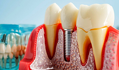
Researchers Solve the Mystery Behind a Billion-Dollar Dental Implant Disease
Researchers have uncovered why a common and costly dental implant infection often resists antibiotics. Dental implants have helped tens of millions of people regain a full set of stable, functional teeth, something traditional dentures [...]
Nanoparticles inspired by lung fluid improve therapies targeting respiratory system
The CIC biomaGUNE Center for Cooperative Research in Biomaterials has developed pulmonary surfactant nanoparticles (the blend of lipids and proteins that line the alveoli and enables breathing), which are encapsulated [...]

Scientists Finally Uncover How a “Forever Chemical” Causes Birth Defects
PFDA, a PFAS “forever chemical,” can cause craniofacial birth defects by disrupting retinoic acid regulation during fetal development, revealing the first clear molecular mechanism behind the link. Researchers have long linked perfluoroalkyl and polyfluoroalkyl substances (PFAS), [...]
Scientists Have Discovered These Deadly Parasites Are Secretly Swapping DNA
Leishmania parasites appear to evolve through widespread genetic exchange, reshaping assumptions about how they adapt and spread. A parasite long thought to spread mostly by cloning itself may be far more genetically dynamic than [...]
Stanford’s Revolutionary New Microscope Reveals Living Cells in Stunning Detail
Stanford researchers have developed a microscope that can show how nanostructures interact inside living cells at the highest resolution achieved so far. The view into living cells just got better. Stanford researchers have merged [...]