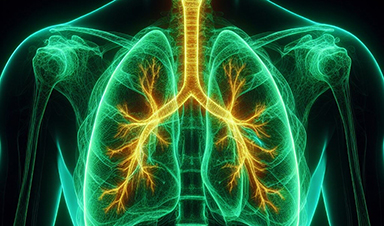
Researchers at National Jewish Health have shown that subtle increases in lung scarring, detected by an artificial intelligence-based tool on CT scans taken one year apart, are associated with disease progression and survival in patients with fibrotic interstitial lung disease. The findings, recently published in the American Journal of Respiratory and Critical Care Medicine, suggest that computer-based image analysis may provide an earlier, more objective way to identify patients at highest risk for worsening disease.
“We found that even small increases in fibrosis over one year signal a higher risk of lung function decline and mortality,” said Matthew Koslow, MD, pulmonologist at National Jewish Health and lead co-author of the study. “For example, patients with a 5% or more increase in fibrosis score showed a greater than two-fold increased risk of death or lung transplant and steeper declines in lung function in the following year compared to patients with stable fibrosis scores. What is especially important is that these changes were strongest in patients with less severe disease at baseline —precisely the group where earlier intervention has the greatest potential to alter the course of disease.”
Fibrotic interstitial lung diseases, which include idiopathic pulmonary fibrosis, or IPF, are a group of chronic, progressive disorders marked by lung scarring that make breathing increasingly difficult. Current tools for predicting progression rely on symptoms, lung function tests and radiologist interpretation of high-resolution CT scans—each of which can be limited by subjectivity or variability, especially when evaluating changes over time.
The new study used a deep learning method called data-driven textural analysis (DTA). Developed by the Quantitative Imaging Laboratory at National Jewish Health, DTA provides a precise measurement of the extent of lung fibrosis on CT scans. Researchers found that increases in DTA fibrosis scores over one year were strongly associated with subsequent lung function decline and a higher risk of death or lung transplant.
“This work demonstrates how quantitative imaging and robust statistical modeling can uncover meaningful patterns in disease progression,” said David Baraghoshi, Ph.D., biostatistician at National Jewish Health and co-first author of the study. “By analyzing changes in fibrosis scores over time and linking them to future outcomes, we were able to show that imaging data can serve as a powerful marker of clinical trajectory.”
The results were validated using data from the Pulmonary Fibrosis Foundation Patient Registry, underscoring the generalizability of the findings.
These insights could have major implications for clinical trials and patient care. Quantitative CT analysis may serve as a meaningful trial endpoint, a tool for selecting patients at highest risk, and a guide for treatment decisions in real-world practice.
More information: Matthew Koslow et al, One-Year Change in Quantitative Computed Tomography Is Associated with Meaningful Outcomes in Fibrotic Lung Disease, American Journal of Respiratory and Critical Care Medicine (2025). DOI: 10.1164/rccm.202503-0535oc
News

Popular Vitamin B3 Supplements May Help Cancer Cells Survive, Scientists Warn
A new study raises important questions about widely used NAD+ supplements, suggesting that compounds often taken to boost energy and support healthy aging may have unintended consequences in cancer treatment. Millions of Americans take [...]

Scientists Discover Cancer Tumors Are “Addicted” to This Common Antioxidant
Cancer cells may be exploiting a common antioxidant as fuel, revealing a potential weakness that future therapies could target. Cancer cells may be tapping into an unexpected energy source: an antioxidant long associated with [...]
Nanotube injector transfers cytoplasmic contents and organelles between living cells safely
Cells are not isolated units; they continuously exchange proteins, genetic material, and even entire organelles with their neighbors. Intercellular transfer influences how tissues develop, respond to stress, and repair damage. In certain cancers, for [...]

CEO of America’s largest public hospital system is ready to replace radiologists with AI
The chief executive of America’s largest public hospital system says he is prepared to start replacing radiologists with artificial intelligence in some circumstances, once the regulatory landscape catches up. Mitchell H. Katz, MD, president [...]

Our books now available worldwide!
Online Sellers other than Amazon, Routledge, and IOPP Indigo Global Health Care Equivalency in the Age of Nanotechnology, Nanomedicine and Artifcial Intelligence Global Health Care Equivalency In The Age Of Nanotechnology, Nanomedicine And Artificial [...]

Study finds higher heart disease risk in long COVID patients
People with long COVID are at increased risk of developing cardiovascular disease, according to a new study from Karolinska Institutet published in eClinicalMedicine. The results show that the risk of conditions such as cardiac arrhythmias [...]
The Corona variant Cicada is here – we know that
Online and on social media, reports are piling up about a new Sars-Cov-2 variant that is currently on the rise: BA.3.2, also known as Cicada. That's what it's all about: The Omicron variant BA.3.2, [...]

A Simple Blood Test Could Predict Dementia Risk 25 Years Early
A single blood marker may quietly signal dementia risk decades in advance. Scientists at the University of California, San Diego, have identified a blood signal that could forecast dementia risk decades before symptoms begin. Their [...]

Sperm Get Lost in Space and Scientists Finally Know Why
Having a baby in space may be far more complicated than expected, as new research shows sperm struggle to find their way in microgravity. Starting a family beyond Earth could be more complicated than [...]
Digital Dementia – Brain fog and disassociation from being chronically online
New medical evidence, featured on 60 Minutes Australia, indicates excessive screen time is causing "digital dementia" in young Australians, with brain scans showing physical shrinkage and damage. Experts warn that high device usage (6-8 hours [...]

A new, highly mutated COVID variant called ‘Cicada’ is spreading in the US.
BA.3.2, a heavily mutated new COVID-19 variant which may be better able to escape immunity from vaccines or prior infection, is now spreading in the United States. Although COVID cases are currently low nationally, [...]

Molecular Manufacturing: The Future of Nanomedicine – New book from NanoappsMedical Inc.
This book explores the revolutionary potential of atomically precise manufacturing technologies to transform global healthcare, as well as practically every other sector across society. This forward-thinking volume examines how envisaged Factory@Home systems might enable the cost-effective [...]

Ancient bacteria strain discovered in ice cave is resistant to some modern antibiotics
In the depths of Scarisoara cave in Romania sits one of the world’s biggest underground glaciers, a monumental slab of ice the size of roughly 40 Olympic swimming pools that began to form around [...]

Scientists Identify “Good” Bacteria That May Prevent Long COVID
According to the WHO, about 6% of people worldwide who get COVID-19, roughly 400 million people, later develop a long-lasting form of the illness. That shows the condition remains a significant public health challenge. In [...]

New book from Nanoappsmedical Inc. – Global Health Care Equivalency
A new book by Frank Boehm, NanoappsMedical Inc. Founder. This groundbreaking volume explores the vision of a Global Health Care Equivalency (GHCE) system powered by artificial intelligence and quantum computing technologies, operating on secure [...]

RNA Recycling Extends Lifespan
Summary: Researchers discovered a biological “trash disposal” mechanism that directly controls how fast we age. While circular RNA has long been known to accumulate in cells as we get older, this study proves for the [...]