In an article recently published in the journal Applied Surface Science, the researchers synthesized green fluorescent carbon dots (G-CDs) from 3,5-diaminobenzoic acid and citric acid. The as-prepared G-CDs were used to target the nucleolus and to carry exogenous DNA molecules into the nucleolus. In addition, the carbon dots (CDs) were used to monitor the levels of nitrite ions (NO2–) and pH in biological cells.
Role of Nucleolus, NO2– and pH in Living Cells
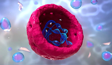
CDs are carbon nanomaterials considered quasispherical particles with a size of fewer than 10 nanometers. Due to their advantages, including outstanding photostability, superior photoluminescence, excellent biocompatibility low toxicity, and easy functionalization, CDs are extensively applied in biological sensing, cell imaging, gene delivery, catalysis, and fingerprints detection.
The nucleolus is located inside the nucleus and it is the site for processing ribosomal RNA (rRNA) transcripts, ribosomal DNA transcription by RNA polymerase I, and ribosome assembly. Monitoring the state of the nucleolus is critical for detecting malignant lesions and developing accurate treatment.
Although CDs were recently applied as fluorescent (FL) labeling reagents for nucleolus staining, their research application as nucleolus-targeted probes is practically unexplored.
Many human diseases are caused by genetic changes inside the nucleolus. Thus, transmitting normal genes with therapeutic efficacy into the nucleolus can exert a therapeutic role or correct gene defects. However, the internalization of naked genetic materials by target cells may be hampered due to phagocyte uptake, susceptibility to serum nuclease, or rapid renal clearance. To this end, various vectors were developed to introduce the gene into the nucleolus. However, these vectors have limitations like high toxicity and low transfection efficiency.
NO2– is used as a preservative in the food industry. Although a moderate amount of NO2– is beneficial to human health, excess presence could convert hemoglobin into methemoglobin in human blood, which may cause hypoxia and interact with secondary amines and amides present in the stomach to produce nitrosamine and may induce cancer and hypertension. Thus, detection of NO2– is critical for disease diagnosis. Similarly, pH plays a vital role in physiological processes, and irregular pH changes lead to various diseases. Measuring intracellular pH is also crucial for disease diagnosis.
G-CDs for Nucleolus Targeting, Gene Delivery, and Biosensing of NO2– and pH
In the present study, the researchers synthesized G-CDs from citric acid and 3,5-diaminobenzoic acid via a single-step hydrothermal method. As-prepared G-CDs showed excellent biocompatibility and low toxicity. The G-CDs were applied to the target nucleolus based on G-CD’s DNA-sensitive properties to carry the exogenous DNA into the nucleolus. Moreover, the G-CDs were used to monitor NO2– and pH in a biological cell.
Research Findings
Transmission electron microscope (TEM) images demonstrated G-CDs’ quasi-spherical structure with favorable dispersion and size distribution in the scope of 1-5 nanometers with an average size of 2.16 ± 0.52 nanometers. Atomic force microscopy revealed that G-CDs’ height range was between 5.2 and 5.1 nanometers.
Fourier transform infrared (FTIR) spectrum and X-ray diffraction patterns inferred the composition and functional groups. G-CD’s XRD pattern showed three characteristic peaks at 281.9, 396.9, and 528.8 electronvolts corresponding to C1s, N1s, and O1s, respectively.
The C1s spectrum illustrated four peaks corroborating the existence of the following bonds, carbon-carbon double (C=C, 284.8 electronvolts), carbon-nitrogen (C-N, 286 electronvolts), acetyl (O=C–C, 287.8 electronvolts) and carbonyl (C=O, 288.9 electronvolts). N1s spectrum decomposed into three peaks corresponding to carbon-nitrogen-carbon (C-N-C, 398.8 electronvolts) and nitrogen-hydrogen (N-H, 400.1 electronvolts). O1s spectrum also decomposed into three peaks of C=O (531.8 electronvolts) and carbon-hydroxyl (C-OH, 533.2 electronvolts), and ether (C-O-C, 533.9 electronvolts)
FTIR spectrum showed absorption bands for N–H and C–H at 2927.3 and 2579.3-centimeter inverse, respectively. The peak at 1690.9-centimeter inverse corroborated the existence of the C=O functional group, those at 1593.9 and 1464.4-centimeter inverse COO– group. Moreover, the peaks at 1394.7, 1334.8, 1210.5, and 1121.1-centimeter inverse correspond to C–H bending vibration, C-N stretching vibrations, O-H and C–O bond, respectively.
G-CD’s complex efficiency for DNA was investigated through an agarose gel electrophoresis assay by observing the movement of DNA. The results showed that free DNA had easy migration to the opposite end, while with the increasing weight ratio of G-CD to DNA, DNA binding with G-CD was gradually blocked, and complete blockage occurred when the ratio reached 50:1. Complete blockage in DNA migration confirms the effective loading of DNA in G-CDs.
Conclusion
To conclude, the researchers used 3,5-diaminobenzoic acid and citric acid as precursors to develop G-CDs via a single-step hydrothermal reaction. The as-prepared G-CDs served the purpose of staining nucleolus, carrying exogenous DNA into nucleolus, and visually monitoring pH and NO2– variations in living cells. Additionally, the team anticipated that the proposed G-CDs were biocompatible gene carriers with low toxicity. Thus, these CDs have good prospects in gene therapy that targets nucleolus.
News

Younger Generations Are Aging Faster – and It May Be Fueling a Surge in Cancer
Younger generations may be aging biologically faster than those before them, and that shift could help explain rising rates of cancer at younger ages. For decades, cancer was viewed largely as a disease of [...]

Using Cannabis Could Raise Your Stroke Risk by 37%, Massive Study Reveals
Large-scale evidence suggests cannabis, cocaine, and amphetamines may directly raise stroke risk, including in younger adults. As recreational drug use becomes increasingly common, researchers are uncovering evidence that its health consequences may extend far beyond [...]

Could Vitamin C Be the Secret to Keeping Your Brain Younger?
Lower vitamin C levels were linked to reduced brain volume and weaker neural connectivity in older adults, suggesting a potential connection between nutrition and brain health. Could a common vitamin help preserve the brain [...]

This Deadly Disease Was Wiping Out Humans 5,500 Years Ago
A new study suggests plague was already a deadly threat 5,500 years ago, striking small hunter-gatherer communities long before cities and agriculture emerged. For centuries, plague has been remembered as the disease that devastated [...]

China closing in but US leads in biotech quality, commercial reach, survey finds
SAN DIEGO, June 22 (Reuters) - China, which now conducts more clinical drug trials, opens new tab than the U.S., still lags in the quality and commercial reach of its biomedical science, according to a recent survey, opens new [...]

New method generates renewable supply of progenitor immune cells
In a paper published in Cell, a USC Stem Cell-led team reports a new way of generating a renewable and expandable supply of the progenitor cells that give rise to macrophages. These immune cells help [...]

Scientists Just Discovered a Cellular Survival System That Was Never Supposed To Exist
A surprising backup pathway allows cells to make a crucial amino acid when their primary machinery fails. For decades, biologists believed cells had only one way to access a molecule they cannot live without. New [...]

Artificial cells gain porous membranes, enabling lab reactions and drug release
Artificial cells created in the laboratory offer a wide range of potential applications. Until now, however, their membranes—unlike those of real cells—have been virtually impermeable. Researchers at the Max Planck Institute for Polymer Research, [...]

Popular Weight-Loss Drugs Like Ozempic Linked to Lower Breast Cancer Risk
Ozempic and similar weight-loss drugs were linked to a striking 30% reduction in breast cancer risk in a study of more than 110,000 women. Popular weight-loss and diabetes medications such as Ozempic, Wegovy, Mounjaro, [...]

Stanford Scientists Discover Explosive New Type of Immune Cell
Scientists studying the remarkable regenerative abilities of planarian flatworms have uncovered a previously unknown type of immune cell with an unusually destructive defense strategy. What if an immune cell could wipe out nearby threats [...]

Big Pharma-backed SonoThera sounds off with $125M series B for bubble-based genetic delivery
Bay Area biotech SonoThera is bubbling to a clinical boil after raising a $125 million series B with the backing of some of the biggest names in pharma. Vida Ventures led the raise, with the venture [...]

Joint initiative of 5 EU countries calls for ‘unified approach’ to pharma framework amid US drug pricing pressure
With drug pricing pressure building from the U.S., a healthcare-focused consortium of five European countries is calling for a “unified approach” to strengthen Europe’s pharmaceutical framework and access to innovative medicines. Belgium, the Netherlands, [...]

Our books now available worldwide!
Online Sellers other than Amazon, Routledge, and IOPP Indigo Global Health Care Equivalency in the Age of Nanotechnology, Nanomedicine and Artifcial Intelligence Global Health Care Equivalency In The Age Of Nanotechnology, Nanomedicine And Artificial [...]

Molecular Manufacturing: The Future of Nanomedicine – New book from NanoappsMedical Inc.
This book explores the revolutionary potential of atomically precise manufacturing technologies to transform global healthcare, as well as practically every other sector across society. This forward-thinking volume examines how envisaged Factory@Home systems might enable the cost-effective [...]

NanoMedical Brain/Cloud Interface – Explorations and Implications. A new book from Frank Boehm
New book from Frank Boehm, NanoappsMedical Inc Founder: This book explores the future hypothetical possibility that the cerebral cortex of the human brain might be seamlessly, safely, and securely connected with the Cloud via [...]

New book from Nanoappsmedical Inc. – Global Health Care Equivalency
A new book by Frank Boehm, NanoappsMedical Inc. Founder. This groundbreaking volume explores the vision of a Global Health Care Equivalency (GHCE) system powered by artificial intelligence and quantum computing technologies, operating on secure [...]