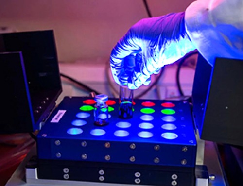
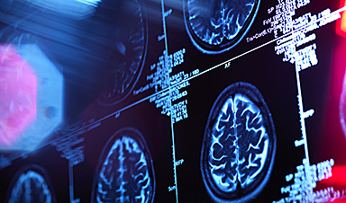
A newly developed imaging method blends ultrasound and photoacoustics to capture both tissue structure and blood-vessel function in 3D.
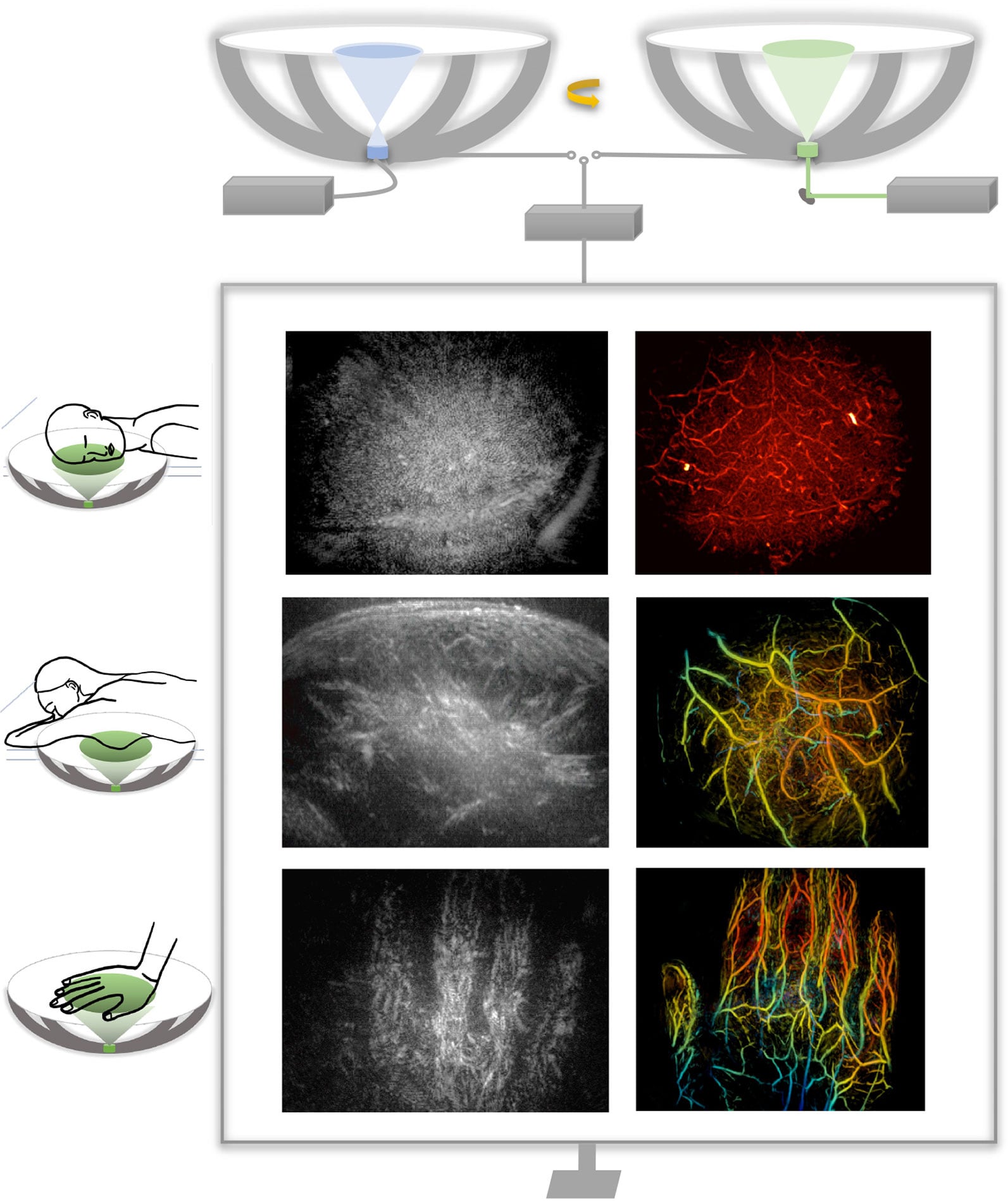
By blending two powerful imaging methods, researchers from Caltech and USC have developed a new way to see inside the human body with unprecedented speed and detail. The technique produces three-dimensional, full-color images that show not only the shape of soft tissues but also how blood vessels are functioning in real time. In early demonstrations, the researchers successfully imaged several different parts of the human body, highlighting the versatility of the approach.
This combined imaging method could significantly improve how doctors detect and study disease. Potential applications include more precise breast tumor imaging, new ways to track nerve damage caused by diabetes, and advanced tools for observing brain structure alongside blood flow. The work suggests a path toward medical scans that are both more informative and easier to repeat over time.
The researchers describe the new technology in a paper published in Nature Biomedical Engineering.
Medical imaging often requires tradeoffs between speed, cost, and the type of information that can be captured. Ultrasound, one of the most widely used techniques, is fast, inexpensive, and well suited for visualizing the structure of tissues. However, it typically provides only two-dimensional views and cannot easily capture a wide area or reveal detailed information about blood chemistry or flow.
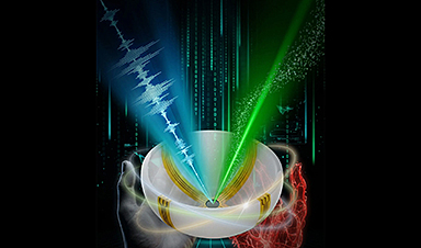
Photoacoustic imaging addresses some of those gaps but introduces others. In this approach, laser light is sent into the body, where it is absorbed by molecules in blood vessels. That absorption generates sound waves that can be detected and translated into images. Because different molecules absorb light in distinct ways, photoacoustic imaging can display blood vessels in optical color—allowing for visualization of how blood moves through arteries and veins. On its own, however, the technique does not provide enough structural detail to fully map surrounding tissues.
Other advanced imaging tools, such as computed tomography (CT) scanning and magnetic resonance imaging (MRI), can deliver detailed views of anatomy, but they come with notable downsides. These methods can be costly, may require contrast agents, sometimes involve exposure to ionizing radiation, or take too long to be practical for frequent monitoring or bedside use.
Combining Ultrasound and Photoacoustics
To overcome these limitations, the researchers developed RUS-PAT (rotational ultrasound tomography, RUST, combined with photoacoustic tomography, PAT). PAT was first developed more than two decades ago by Lihong Wang, the Bren Professor of Medical Engineering and Electrical Engineering and the Andrew and Peggy Cherng Medical Engineering Leadership Chair at Caltech.
In PAT, molecules that absorb light respond to short laser pulses by vibrating, which generates acoustic signals. These signals can then be detected and processed to form detailed, high-resolution images.
Wang, who is also the executive officer for medical engineering at Caltech, says his group's aim with the current work was to combine the benefits of PAT with ultrasound. "But it's not like one plus one," he says. "We needed to find an optimal way of combining the two technologies."
Ultrasound typically uses many transducers to both generate and receive ultrasound waves, and combining this process directly with PAT would be too complex and expensive for widespread use. PAT, meanwhile, only requires the detection of ultrasound, and that gave Wang an idea. "I thought, 'Wait, can we just mimic light excitation of ultrasound waves in photoacoustic tomography, but do it ultrasonically?'" PAT allows laser light to diffuse within the tissue, ultimately triggering the production of measurable ultrasound waves. Similarly, Wang figured, they could use a single wide-field ultrasound transducer to broadcast an ultrasonic wave broadly into the tissue.
They could then use the same detectors to measure the resulting waves for both modalities. In the new system, a small number of arc-shaped detectors are rotated around a central point, allowing it to behave like a full hemispheric detector but at a fraction of the complexity and cost.
Demonstrated Clinical Potential
"The novel combination of acoustic and photoacoustic techniques addresses many of the key limitations of widely used medical-imaging techniques in current clinical practice, and, importantly, the feasibility for human application has been demonstrated here in multiple contexts," says Dr. Charles Y. Liu, an author of the paper who is a visiting associate in biology and biological engineering at Caltech. Liu is also a professor at the Keck School of Medicine of USC, director of USC's Neurorestoration Center, and chair of neurosurgery at the Rancho Los Amigos National Rehabilitation Center.
The RUS-PAT technique could potentially be used in any region of the body to which light can be delivered, and for applications where clinicians or researchers would benefit from the synergistic imaging of both the morphology and color-related function. For example, RUS-PAT could improve breast-tumor imaging, giving physicians the ability to know a tumor's exact location and surroundings as well as its pathology and physiology. It could also help doctors monitor the nerve damage caused by diabetic neuropathy by providing an all-in-one way to monitor oxygen supply along with morphology. Wang says the technique could also be useful in brain imaging, allowing scientists to observe the structural details of the brain while also being able to observe hemodynamics.
Currently, the system can scan to a depth of about 4 centimeters. Light can also be delivered endoscopically, potentially making deeper tissues accessible to the new technology. A RUS-PAT scan can be performed in less than one minute.
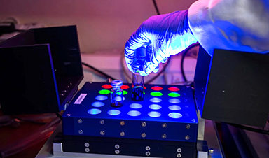
The current setup involves a scanning system with ultrasound transducers and laser housed underneath a bed. It has been demonstrated on human volunteers and patients and is in the early stages of translational development.
Reference: "Rotational ultrasound and photoacoustic tomography of the human body" by Yang Zhang, Shuai Na, Jonathan J. Russin, Karteekeya Sastry, Li Lin, Junfu Zheng, Yilin Luo, Xin Tong, Yujin An, Peng Hu, Konstantin Maslov, Tze-Woei Tan, Charles Y. Liu and Lihong V. Wang, 16 January 2026, Nature Biomedical Engineering.
DOI: 10.1038/s41551-025-01603-5
The work was supported by funding from the National Institutes of Health.
News
A Cambridge Lab Mistake Reveals a Powerful New Way to Modify Drug Molecules
A surprising lab discovery reveals a light-powered way to tweak complex drugs faster, cleaner, and later in development. Researchers at the University of Cambridge have created a new technique for altering complex drug molecules [...]

New book from NanoappsMedical Inc – Molecular Manufacturing: The Future of Nanomedicine
This book explores the revolutionary potential of atomically precise manufacturing technologies to transform global healthcare, as well as practically every other sector across society. This forward-thinking volume examines how envisaged Factory@Home systems might enable the cost-effective [...]
Scientists Discover Simple Saliva Test That Reveals Hidden Diabetes Risk
Researchers have identified a potential new way to assess metabolic health using saliva instead of blood. High insulin levels in the blood, known as hyperinsulinemia, can reveal metabolic problems long before obvious symptoms appear. It is [...]

One Nasal Spray Could Protect Against COVID, Flu, Pneumonia, and More
A single nasal spray vaccine may one day protect against viruses, pneumonia, and even allergies. For decades, scientists have dreamed of creating a universal vaccine capable of protecting against many different pathogens. The idea [...]
New AI Model Predicts Cancer Spread With Incredible Accuracy
Scientists have developed an AI system that analyzes complex gene-expression signatures to estimate the likelihood that a tumor will spread. Why do some tumors spread throughout the body while others remain confined to their [...]

Scientists Discover DNA “Flips” That Supercharge Evolution
In Lake Malawi, hundreds of species of cichlid fish have evolved with astonishing speed, offering scientists a rare opportunity to study how biodiversity arises. Researchers have identified segments of “flipped” DNA that may allow fish to adapt rapidly [...]

Our books now available worldwide!
Online Sellers other than Amazon, Routledge, and IOPP Indigo Global Health Care Equivalency in the Age of Nanotechnology, Nanomedicine and Artifcial Intelligence Global Health Care Equivalency In The Age Of Nanotechnology, Nanomedicine And Artificial [...]
Scientists Discover Why Some COVID Survivors Still Can’t Taste Food Years Later
A new study provides the first direct biological evidence explaining why some people continue to experience taste loss long after recovering from COVID-19. Researchers have uncovered specific biological changes in taste buds that could help [...]
Catching COVID significantly raises the risk of developing kidney disease, researchers find
Catching Covid significantly raises the risk of developing deadly kidney disease, research has shown. The virus was found to increase the chances that patients will develop the incurable condition by around 50 per cent. [...]

New Toothpaste Stops Gum Disease Without Harming Healthy Bacteria
Researchers have developed a targeted approach to combat periodontitis without disrupting the natural balance of the oral microbiome. The innovation could reshape how gum disease is treated while preserving beneficial bacteria. The human mouth [...]

Plastic Without End: Are We Polluting the Planet for Eternity?
The Kunming Montreal Global Biodiversity Framework calls for the elimination of plastic pollution by 2030. If that goal has been clearly set, why have meaningful measures that create real change still not been implemented? [...]
Scientists Rewire Natural Killer Cells To Attack Cancer Faster and Harder
Researchers tested new CAR designs in NK-92 cells and found the modified cells killed tumor cells more effectively, showing stronger anti-cancer activity. Researchers at the Ribeirão Preto Blood Center and the Center for Cell-Based [...]
New “Cellular” Target Could Transform How We Treat Alzheimer’s Disease
A new study from researchers highlights an unexpected player in Alzheimer’s disease: aging astrocytes. Senescent astrocytes have been identified as a major contributor to Alzheimer’s progression. The cells lose protective functions and fuel inflammation, particularly in [...]
Treating a Common Dental Infection… Effects That Extend Far Beyond the Mouth
Successful root canal treatment may help lower inflammation associated with heart disease and improve blood sugar and cholesterol levels. Treating an infected tooth with a successful root canal procedure may do more than relieve [...]
Microplastics found in prostate tumors in small study
In a new study, researchers found microplastics deep inside prostate cancer tumors, raising more questions about the role the ubiquitous pollutants play in public health. The findings — which come from a small study of 10 [...]
All blue-eyed people have this one thing in common
All Blue-Eyed People Have This One Thing In Common Blue Eyes Aren’t Random—Research Traces Them Back to One Prehistoric Human It sounds like a myth at first — something you’d hear in a folklore [...]