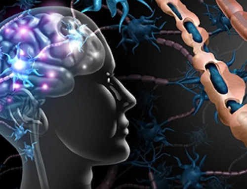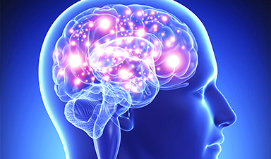
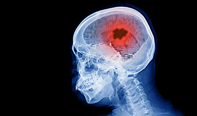
Scientists can depend on a formidable tool — light — when it comes to recording and stimulating brain activity. An international team of researchers, directed by Istituto Italiano di Tecnologia (Italian Institute of Technology/IIT), has created nanometric light modulators that, built on a micrometric optical fiber, make the fiber capable of analyzing neuronal tissue in deep regions of the brain.
The new method, reported in Advanced Optical Materials and presented on the journal’s front cover, lays the foundation for a ground-breaking type of minimally invasive neural probe that can be used to examine the central nervous system. The nanomodulators will be used to explore particular brain diseases, including epilepsy and brain tumors.
The research was performed by IIT in partnership with the University of Salento (Italy), the Politecnico of Bari (Italy), the Consejo Superior de Investigaciones Cientificas (CSIC, Spain) and the Centro National de Investigaciones Oncologicas (CNIO, Spain).
In Italy, the interdisciplinary team was focused on obtaining micrometric structures that are capable of examining neuronal tissue in a thorough way using light, i.e., through the integration of optical nanomodulators.
To achieve this, researchers integrated expertise in nanoscale fabrication and biomedical neuro-engineering, so as to manipulate the physics of surface plasmon polaritons and develop an investigative tool that adjusts and amplifies the way light can excite and monitor particular areas of the brain.
They began from a tapered optical fiber, thinner than a hair, and then they fitted it with nanostructures that resonate in reaction to a light stimulus added by the fiber itself into the deep brain regions.
The nanostructures were developed by coating the microscopic tip of the probe with a thin layer of gold. Then, using a gallium ion beam as a chisel, they fashioned a grid of nanoscopic optical elements, comprising 100 nm thin lines, whose features were confirmed in a series of microscopy and optical spectroscopy experiments.
As a result of this production technique, it was possible to acquire a tool that allows both the probe light beam modulation and the local electric field acting on surfaces comparable to the size of brain cells to be regulated. Scientists may be able to then explore the interaction between the light beam and neuronal structures, even in the deepest areas of the brain.
The possibility of developing such implantable plasmonic systems provides a new perspective in the research of the central nervous system: the amplification created by the nanostructures is meant to be an efficient tool for sensing the biochemical and cellular structure changes underlying the source of numerous neural disorders.
Thus, the part of the international team working out of Spain is concentrating on the application, it may have. Experimental scientists at CSIC led by Liset M de la Prida are aiming to apply these probes in the exploration of post-traumatic epilepsy and neurodegenerative diseases, for example, Alzheimer’s disease.
While the Brain Metastasis Group directed by Manuel Valiente at the CNIO will explore the use of this new technology to differentiate primary from metastatic tumors, whose treatments are diverse, as well as the use of light to permeabilize the blood-brain barrier, permitting anti-tumor drugs to travel via the vascular barrier.
News
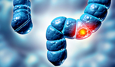
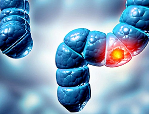
Why More People in Their 30s Are Suddenly Getting Colon Cancer
A major Swiss study found that colorectal cancer is becoming increasingly common in adults under 50, even as rates decline in older age groups. Researchers in Switzerland have identified a concerning trend: while colorectal [...]
Researchers Compare MS Models to Human Tissue in Search for Better Therapies
Researchers identified key differences between two widely used multiple sclerosis models, showing how each can better study myelin damage, immune responses, and repair. The findings may improve efforts to develop treatments that restore lost [...]
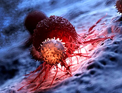
Scientists Discover Genetic “Off Switch” That Supercharges CAR T Cells Against Cancer
A new study reveals a possible way to make CAR T-cell therapy more durable and effective by targeting a single gene-regulating protein. CAR T-cell therapy is widely seen as a breakthrough in personalized cancer [...]
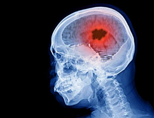
New Vitamin B12-Based Therapy Could Change How Brain Cancer Is Treated
Researchers have identified a vitamin B12–based compound that appears capable of crossing the blood–brain barrier and selectively accumulating in glioblastoma tissue. For decades, one of the biggest problems in brain cancer treatment has had [...]

Simple Fiber Supplement Cuts Knee Arthritis Pain in Just 6 Weeks, Study Finds
A daily inulin supplement may help reduce knee osteoarthritis pain while revealing a possible link between gut health, muscle function, and pain sensitivity. For millions of people living with knee osteoarthritis, managing chronic pain [...]

This Common Vitamin May Help Stop Prediabetes From Turning Into Diabetes
Vitamin D may help prevent type 2 diabetes in people with specific genetic variations, offering a possible path toward personalized diabetes prevention. More than 40% of U.S. adults have prediabetes, a condition in which [...]

Ebola, hantavirus: Is the world prepared for the next pandemic?
Funding cuts to health research and a growing antivaccine movement are making it harder than ever to respond to viruses. The World Health Organization (WHO) has declared that an Ebola outbreak in Uganda and [...]

May 2026 Healthcare News and Trends: Market Signals That Matter
Artificial intelligence is dominating headlines, telehealth has settled into a new normal, and digital health continues to promise transformation. However, much of what is being discussed in healthcare today reflects potential rather than reality. [...]
Scientists Rewire Donor Stem Cells To Outsmart Aggressive Blood Cancers
Researchers have tested a gene-edited stem cell transplant designed to shield healthy blood-forming cells from powerful cancer-targeting immunotherapies. For patients with highly aggressive blood cancers, stem cell transplantation can offer a rare chance at [...]

Recent Digital Health Trends, Insights and News – May 2026
Last month marked continued progress as digital health moves into its next phase — from AI expanding into drug discovery and core infrastructure to new federal pathways accelerating device access and home-based care. Together, [...]
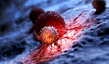
Cancer Mystery Solved: Scientists Discover How Melanoma Becomes “Immortal”
Scientists have uncovered a previously overlooked mechanism that may help melanoma cells become effectively “immortal.” Cancer cells face a major problem before they can become deadly: They have to figure out how to stop [...]
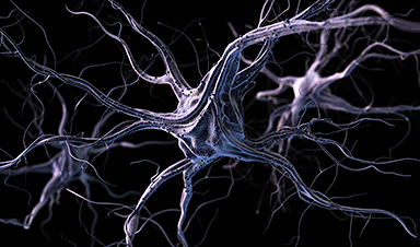
How Visual Neurons Organize Thousands of Synaptic Inputs
Summary: A new study uncovered the organizational rules that determine how neurons in the primary visual cortex process information. By imaging both the cell bodies (soma) and the individual synapses (on dendritic spines) of [...]
Scientists Just Found a Surprising Way To Destroy “Forever Chemicals”
Scientists have uncovered a new mechanism that may help break down highly persistent PFAS pollutants. PFAS have earned the nickname “forever chemicals” for a reason. These industrial compounds are so chemically durable that they [...]
Scientists Discover Cheap Material That Kills Deadly Superbugs
A new sulfur-rich antimicrobial polymer shows strong effectiveness against fungal and bacterial pathogens and may offer an affordable solution to antimicrobial resistance. Antimicrobial resistance is creating growing challenges for both healthcare and food production, [...]

What to Know About Cicada, or BA.3.2, the Latest SARS-CoV-2 Variant Under Monitoring
Like periodical cicadas, the insects for which it is nicknamed, SARS-CoV-2 Omicron subvariant BA.3.2 is only just beginning to emerge after lying low for an extended period since it first appeared. Although it was [...]
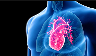
Scientists Say This Simple Supplement May Actually Reverse Heart Disease
Scientists in Japan say a common supplement may actually help “unclog” certain diseased heart arteries from the inside out. A simple food supplement sold in Japan may have helped reverse a dangerous form of [...]