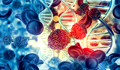
Researchers in Japan found that although the Alzheimer's drug lecanemab successfully removes amyloid plaques from the brain, it does not restore the brain's waste-clearing system within the first few months of treatment.
The study suggests that by the time symptoms appear, damage to nerve cells and the glymphatic system is already well established, making short-term recovery unlikely.
Lecanemab's Surprising Limits in Alzheimer's Treatment
Researchers at Osaka Metropolitan University in Japan, led by graduate student Tatsushi Oura and Dr. Hiroyuki Tatekawa, reported that lecanemab, a drug designed to clear amyloid plaques from the brain, does not improve the brain's waste removal system in the early stages after treatment.
Their results indicate that the nerves of Alzheimer's disease (AD) patients have already sustained considerable damage, and that this waste-clearing function does not rebound quickly. The findings point to the need for treatments that target several biological problems at the same time.
The Complex Web of Alzheimer's Disease Mechanisms
The researchers' results add another piece to the long and complicated effort to understand how AD develops. Although it is the most widespread neurodegenerative disorder, it remains difficult to treat because many different factors contribute to its progression.
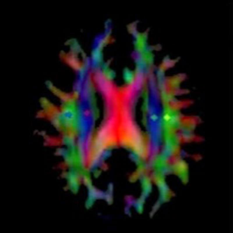
A major driver of nerve cell damage in AD is the accumulation of amyloid-β (Aβ) in the brain. In people without the disease, a network known as the glymphatic system circulates cerebrospinal fluid along the spaces surrounding arteries and into brain tissue. There, this fluid mixes with interstitial fluid to help remove metabolic waste products, including Aβ. The name "glymphatic system" comes from the involvement of glial cells in this process.
In AD, however, Aβ builds up and causes arteries to stiffen. This reduces the movement of fluid between the brain and the cerebrospinal fluid, which disrupts waste removal and leads to a series of harmful changes that produce AD symptoms.
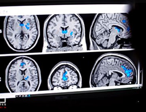
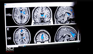
Testing Lecanemab's Effects on Brain Clearance
Lecanemab, which was recently approved as a therapeutic option, is intended to lower Aβ levels. To study its effects, the Osaka Metropolitan University team examined the glymphatic system in patients both before and after lecanemab treatment, using the DTI-ALPS index to measure changes.
They found no meaningful difference in this index when comparing pre-treatment results with measurements taken three months after therapy.
What This Means for Future Alzheimer's Treatments
Based on these observations, the researchers concluded that while anti-amyloid drugs can reduce plaque buildup and slow additional cognitive decline, they may not be able to restore functions already lost. By the time symptoms appear, many patients likely have long-standing neuronal damage and glymphatic impairment that are difficult to reverse. The results emphasize how many interconnected factors drive AD and how few of them can be quickly repaired.
"Even when Aβ is reduced by lecanemab, impairment of the glymphatic system may not recover within the short-term," Oura said. "In the future, we want to look at factors like age, the stage of the disease, and degree of lesions in the white matter to further understand the relationship between changes in the glymphatic system due to lecanemab treatment and the outcome of treatment. This will help understand the best way to administer treatment to patients."
Reference: "Unchanged Early Diffusion Tensor Imaging Along Perivascular Space Index After Amyloid-Targeting Disease-Modifying Therapy in Alzheimer's Disease: A Preliminary Study" by Tatsushi Oura, Hiroyuki Tatekawa, Akitoshi Takeda, Ayako Omori, Natsuko Atsukawa, Shu Matsushita, Daisuke Horiuchi, Hirotaka Takita, Taro Shimono, Daiju Ueda, Yoshiaki Itoh and Yukio Miki, 8 September 2025, Journal of Magnetic Resonance Imaging.
DOI: 10.1002/jmri.70118
The study was published in Journal of Magnetic Resonance Imaging.
News

The Corona variant Cicada is here – we know that
Online and on social media, reports are piling up about a new Sars-Cov-2 variant that is currently on the rise: BA.3.2, also known as Cicada. That's what it's all about: The Omicron variant BA.3.2, [...]

A Simple Blood Test Could Predict Dementia Risk 25 Years Early
A single blood marker may quietly signal dementia risk decades in advance. Scientists at the University of California, San Diego, have identified a blood signal that could forecast dementia risk decades before symptoms begin. Their [...]

Sperm Get Lost in Space and Scientists Finally Know Why
Having a baby in space may be far more complicated than expected, as new research shows sperm struggle to find their way in microgravity. Starting a family beyond Earth could be more complicated than [...]
Digital Dementia – Brain fog and disassociation from being chronically online
New medical evidence, featured on 60 Minutes Australia, indicates excessive screen time is causing "digital dementia" in young Australians, with brain scans showing physical shrinkage and damage. Experts warn that high device usage (6-8 hours [...]

A new, highly mutated COVID variant called ‘Cicada’ is spreading in the US.
BA.3.2, a heavily mutated new COVID-19 variant which may be better able to escape immunity from vaccines or prior infection, is now spreading in the United States. Although COVID cases are currently low nationally, [...]

Molecular Manufacturing: The Future of Nanomedicine – New book from NanoappsMedical Inc.
This book explores the revolutionary potential of atomically precise manufacturing technologies to transform global healthcare, as well as practically every other sector across society. This forward-thinking volume examines how envisaged Factory@Home systems might enable the cost-effective [...]

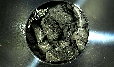
Ancient bacteria strain discovered in ice cave is resistant to some modern antibiotics
In the depths of Scarisoara cave in Romania sits one of the world’s biggest underground glaciers, a monumental slab of ice the size of roughly 40 Olympic swimming pools that began to form around [...]

Scientists Identify “Good” Bacteria That May Prevent Long COVID
According to the WHO, about 6% of people worldwide who get COVID-19, roughly 400 million people, later develop a long-lasting form of the illness. That shows the condition remains a significant public health challenge. In [...]

New book from Nanoappsmedical Inc. – Global Health Care Equivalency
A new book by Frank Boehm, NanoappsMedical Inc. Founder. This groundbreaking volume explores the vision of a Global Health Care Equivalency (GHCE) system powered by artificial intelligence and quantum computing technologies, operating on secure [...]
RNA Recycling Extends Lifespan
Summary: Researchers discovered a biological “trash disposal” mechanism that directly controls how fast we age. While circular RNA has long been known to accumulate in cells as we get older, this study proves for the [...]
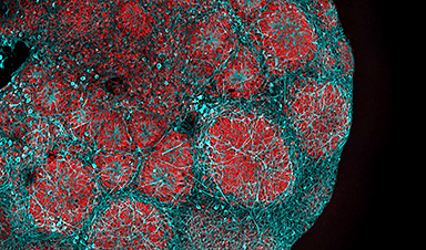
Cancer’s Deadly Paradox: How Tumors Break Their Own DNA To Keep Growing
Cancer’s strongest gene switches push DNA into damaging overdrive, creating repeated breaks and repairs that may fuel tumor evolution while exposing possible therapeutic weak spots. A new study indicates that cancer can harm its own genetic [...]

NanoMedical Brain/Cloud Interface – Explorations and Implications. A new book from Frank Boehm
New book from Frank Boehm, NanoappsMedical Inc Founder: This book explores the future hypothetical possibility that the cerebral cortex of the human brain might be seamlessly, safely, and securely connected with the Cloud via [...]

Our books now available worldwide!
Online Sellers other than Amazon, Routledge, and IOPP Indigo Global Health Care Equivalency in the Age of Nanotechnology, Nanomedicine and Artifcial Intelligence Global Health Care Equivalency In The Age Of Nanotechnology, Nanomedicine And Artificial [...]
Ryugu asteroid samples contain all DNA and RNA building blocks, bolstering origin-of-life theories
All the essential ingredients to make the DNA and RNA underpinning life on Earth have been discovered in samples collected from the asteroid Ryugu, scientists said Monday. The discovery comes after these building blocks [...]

Is Berberine Really a “Natural Ozempic”?
Often labeled a “natural Ozempic,” berberine is widely discussed as a metabolic aid. Yet research suggests its influence may lie deeper. In recent years, berberine has gained significant attention as a supposed “natural way” [...]
Viagra Ingredient Shows Promise for Rare Childhood Brain Disease in Surprising Study
A rare childhood disease with no approved treatment may have an unexpected new therapeutic candidate. Sildenafil, the active ingredient also sold under the brand name Viagra, may help reduce symptoms in people with Leigh [...]