Atomic force microscopy, or AFM, is a widely used technique that can quantitatively map material surfaces in three dimensions, but its accuracy is limited by the size of the microscope’s probe. A new AI technique overcomes this limitation and allows microscopes to resolve material features smaller than the probe’s tip.
“Accurate surface height profiles are crucial to nanoelectronics development as well as scientific studies of material and biological systems, and AFM is a key technique that can measure profiles noninvasively,” said Yingjie Zhang, a U. of I. materials science & engineering professor and the project lead. “We’ve demonstrated how to be even more precise and see things that are even smaller, and we’ve shown how AI can be leveraged to overcome a seemingly insurmountable limitation.”
Often, microscopy techniques can only provide two-dimensional images, essentially providing researchers with aerial photographs of material surfaces. AFM provides full topographical maps accurately showing the height profiles of the surface features. These three-dimensional images are obtained by moving a probe across the material’s surface and measuring its vertical deflection.
If surface features approach the size of the probe’s tip—about 10 nanometers—then they cannot be resolved by the microscope because the probe becomes too large to “feel out” the features. Microscopists have been aware of this limitation for decades, but the U. of I. researchers are the first to give a deterministic solution.
“We turned to AI and deep learning because we wanted to get the height profile—the exact roughness—without the inherent limitations of more conventional mathematical methods,” said Lalith Bonagiri, a graduate student in Zhang’s group and the study’s lead author.
The researchers developed a deep learning algorithm with an encoder-decoder framework. It first “encodes” raw AFM images by decomposing them into abstract features. After the feature representation is manipulated to remove the undesired effects, it is then “decoded” back into a recognizable image.
To train the algorithm, the researchers generated artificial images of three-dimensional structures and simulated their AFM readouts. The algorithm was then constructed to transform the simulated AFM images with probe-size effects and extract the underlying features.
“We actually had to do something nonstandard to achieve this,” Bonagiri said. “The first step of typical AI image processing is to rescale the brightness and contrast of the images against some standard to simplify comparisons. In our case, though, the absolute brightness and contrast is the part that’s meaningful, so we had to forgo that first step. That made the problem much more challenging.”
To test their algorithm, the researchers synthesized gold and palladium nanoparticles with known dimensions on a silicon host. The algorithm successfully removed the probe tip effects and correctly identified the three-dimensional features of the nanoparticles.
“We’ve given a proof-of-concept and shown how to use AI to significantly improve AFM images, but this work is only the beginning,” Zhang said. “As with all AI algorithms, we can improve it by training it on more and better data, but the path forward is clear.”
More information: Lalith Krishna Samanth Bonagiri et al, Precise Surface Profiling at the Nanoscale Enabled by Deep Learning, Nano Letters (2024). DOI: 10.1021/acs.nanolett.3c04712
News

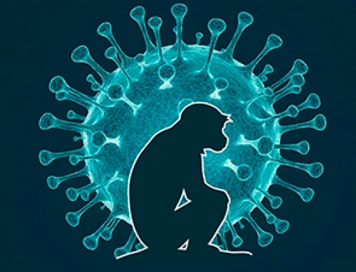
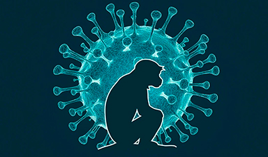
A Deadly Ebola-Like Virus Is Spreading. Are We Ready?
BU virologist Nancy Sullivan says the Bundibugyo outbreak in the Democratic Republic of the Congo underscores the need for broader outbreak preparedness. The death of a nurse marked the moment health officials recognized that [...]
Why Most Animal Viruses Never Become Human Pandemics
From receptor mismatch to risky human-animal interfaces, this article explains why spillover is common but true pandemic emergence remains rare. Introduction Humans are constantly exposed to animal viruses through farming, wildlife contact, and the [...]

Stem cell organoids repair heart microvessels in coronary artery disease models
A Stanford University team has shown that vascular organoids derived from human stem cells can repair the heart’s microvessel network in pigs with ischaemic heart disease – a proof-of-concept advancement that could open new therapeutic [...]

Goodbye GP waiting rooms, hello prevention at home
Prevention is suddenly everywhere in NHS reform. The recent £340m community pharmacy deal is moving more services onto the high street. Community Diagnostic Centres are being expanded, and the Neighbourhood Health Framework sets out [...]
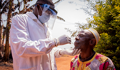
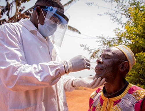
Ebola control is weakened by mistrust and cultural insensitivity
Effective response depends on cooperation with communities and frontline workers, writes Zaeem ul Haq The current Bundibugyo Ebola outbreak in the Democratic Republic of the Congo (DRC) and Uganda is exposing dangerous gaps in [...]

Building the Brain Requires Millions of Dangerous DNA Breaks
Scientists discovered that building a healthy brain involves an unexpected step: young neurons routinely break and rapidly repair their own DNA. As the brain develops, newly formed nerve cells must travel through tightly packed tissue [...]

One Tiny Change May Explain How Viruses Jump From Bats to Humans
Scientists found that one tiny genetic change may determine whether a bat virus stays in bats or becomes a human threat. Most infectious disease outbreaks begin when a virus or other pathogen crosses from animals into [...]

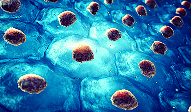
Scientists Discover 250+ Genes That Could Lead to New Ways To Prevent Melanoma
The world’s largest study of mole genetics identified hundreds of genes tied to melanoma risk, uncovering potential new drug targets and paving the way for more accurate melanoma screening and prevention. Researchers at QIMR [...]
Breakthrough Diabetes Treatment Reprograms the Immune System
An engineered stem cell therapy reversed new-onset Type 1 diabetes in mice by shifting the immune system away from attacking insulin-producing cells. For more than a century, people with Type 1 diabetes have relied [...]
Taking the world’s temperature: WHO chief spotlights global health emergencies
Taking the world’s temperature on pressing health matters, WHO Director-General Tedros Adhanom Ghebreyesus provided the latest on current global challenges - and successes when it comes to international cooperation. “The outbreaks of hantavirus, Ebola and Marburg all show [...]
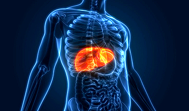
Scientists Create Tiny “Mini Livers” That Could One Day Replace Liver Transplants
Engineered tissue grafts could help perform key liver functions and benefit thousands of people living with liver failure. The liver is one of the body’s hardest-working organs, carrying out hundreds of vital jobs, from [...]

NanoMedical Brain/Cloud Interface – Explorations and Implications. A new book from Frank Boehm
New book from Frank Boehm, NanoappsMedical Inc Founder: This book explores the future hypothetical possibility that the cerebral cortex of the human brain might be seamlessly, safely, and securely connected with the Cloud via [...]
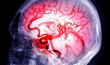
Scientists Discover Surprising Way To Help the Brain Recover After Stroke
A new study suggests that strengthening the body’s natural circadian rhythms may help the brain recover after stroke, even when treatment begins days after the injury. Every year, millions of people survive a stroke, [...]

Our books now available worldwide!
Online Sellers other than Amazon, Routledge, and IOPP Indigo Global Health Care Equivalency in the Age of Nanotechnology, Nanomedicine and Artifcial Intelligence Global Health Care Equivalency In The Age Of Nanotechnology, Nanomedicine And Artificial [...]

Younger Generations Are Aging Faster – and It May Be Fueling a Surge in Cancer
Younger generations may be aging biologically faster than those before them, and that shift could help explain rising rates of cancer at younger ages. For decades, cancer was viewed largely as a disease of [...]

Using Cannabis Could Raise Your Stroke Risk by 37%, Massive Study Reveals
Large-scale evidence suggests cannabis, cocaine, and amphetamines may directly raise stroke risk, including in younger adults. As recreational drug use becomes increasingly common, researchers are uncovering evidence that its health consequences may extend far beyond [...]