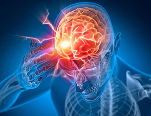
Malignant brain tumors are cancerous growth in the brain with the possibility of spreading to other parts of the central nervous system (CNS). Brain tumors are highly invasive and have devastating consequences, poor prognosis, and low survival rates.
Recently, the noninvasive near-infrared fluorescence and photoacoustic imaging techniques have demonstrated a bright scope in brain tumor diagnosis with high spatiotemporal resolution, large penetration depth, and good sensitivity. Hence, these imaging techniques have been used to guide real-time brain tumor therapy with high efficiency and accuracy.
An article published in the journal Advanced Drug Delivery Reviews presented a summary of state-of-art near-infrared contrast agents (CAs) and optical imaging-guided therapies for brain tumor imaging. Finally, the opportunities and challenges involved in the use of CAs and nanotheranostics for future clinical translation were discussed.
Near-Infrared Fluorescence Imaging of Brain Tumors
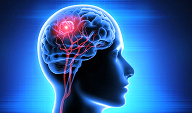
The brain tumor is an intracranial neoplasm in the brain or the central spinal canal. An abnormal and uncontrolled cell division, usually in the brain, involving neurons or glial cells or occasionally in the lymphatic tissue, blood vessels, and others, is the main leading cause of the formation of primary brain tumors. The majority of the brain tumors in adults are secondary or metastatic tumors, that is, cancers primarily located in other organs and may spread to the brain and create brain tumors.
Diagnosis of brain tumors is the critical step for effective curing of the disease, and it vividly relies on the advancements in molecular imaging technology. Although various imaging modalities, including magnetic resonance imaging (MRI), positron emission tomography (PET) imaging, and computed tomography (CT) imaging were investigated for the detection of brain tumors, these modalities possess several limitations.
In this regard, fluorescence imaging is a method that relies on fluorescence, absorption, bioluminescence, and reflectance resulting from various fluorescent nanomaterials used for visualizing the brain microstructures and monitoring the tumor progression.
Nevertheless, the effectiveness and safety of fluorescence imaging, using visible light for fluorescence imaging, displayed limited penetration of the light and resulted in decreased light-tissue interactions. Light in the near-infrared range (wavelength: 650–900 nanometers) has several advantages over visible-range light, including deeper tissue penetration due to less absorption by hemoglobin and water and less autofluorescence from surrounding tissues. Consequently, a new scope has emerged for near-infrared fluorescence imaging for detecting and treating brain tumors.
Near-infrared fluorescence imaging of brain tumors is a growing field for preclinical and clinical applications in clinical management due to its advantageous features, including a high spatial resolution, portability, real-time display, and detailed molecular profiling with the multiplexed use of fluorescent probes.
Similarly, near-infrared photoacoustic imaging (PAI) is a noninvasive imaging technique that involves acoustic waves as the emission source. It combines the advantages of ultrasonic and optical energies to realize biological imaging with deep tissue penetration depth. Thus, making PAI a promising diagnosis technique.
Near Infrared Optical Imaging-Guided Treatment of Brain Tumors
Near-infrared optical imaging-guided brain tumor therapy has been used in theranostics. Various therapeutic functions have been demonstrated with the assistance of near-infrared fluorescence imaging and PAI.
Despite encouraging progress, several obstacles remain in transitioning optical imaging techniques to clinical applications. As safety is the primary concern for the clinical translation of nanomedicine, nanotheranostics and nanoprobes are subjected to various surface modifications using biocompatible polymers to reduce their toxicities.
Near-infrared fluorescence imaging and PAI have vividly helped in the diagnosis of cancer. However, the presence of the skull and scalp hampers the quality of light and the imaging of brain tumors. This suggests that the second near-infrared region (NIR-II) can increase tissue penetration and depth while decreasing light scattering and enhancing the signal-to-background ratio. Consequently, deep-seated brain tumors can also be diagnosed.
CAs with excited-state intramolecular motion have good prospects for fine-tuning the balance between nonradiative and radiative decay in dual-mode near-infrared fluorescence imaging or PAI. Near-infrared fluorescence imaging CAs, including organic dyes, semiconducting polymer dots, aggregation-induced emission luminous, and inorganic (quantum dots and rare-earth nanoparticles) probes, have been investigated for the diagnosis of brain tumors.
Conclusion
Overall, multimodal optical imaging techniques can decrease misdiagnosis rates of brain tumors and provide functional and anatomical information. Moreover, theranostic agent-based imaging guidance and combination treatment can enhance the treatment outcomes of brain tumors and reduce side effects.
Furthermore, nanoparticles less than 5 nanometers in size are favorable for renal excretion and reduce toxicity during treatment. Thus, the fabrication of biodegradable nanomaterials followed by toxicity evaluations can increase the clinical success of treatment.
In the case of brain tumors, the blood-brain barrier restricts the entry of nanoagents into brain tissues. In this regard, peptides with targeting abilities and focused ultrasound (FUS) assistance can help deliver nanoagents to brain tumors for diagnosis and therapy.

Younger Generations Are Aging Faster – and It May Be Fueling a Surge in Cancer
Younger generations may be aging biologically faster than those before them, and that shift could help explain rising rates of cancer at younger ages. For decades, cancer was viewed largely as a disease of [...]

Using Cannabis Could Raise Your Stroke Risk by 37%, Massive Study Reveals
Large-scale evidence suggests cannabis, cocaine, and amphetamines may directly raise stroke risk, including in younger adults. As recreational drug use becomes increasingly common, researchers are uncovering evidence that its health consequences may extend far beyond [...]

Could Vitamin C Be the Secret to Keeping Your Brain Younger?
Lower vitamin C levels were linked to reduced brain volume and weaker neural connectivity in older adults, suggesting a potential connection between nutrition and brain health. Could a common vitamin help preserve the brain [...]

This Deadly Disease Was Wiping Out Humans 5,500 Years Ago
A new study suggests plague was already a deadly threat 5,500 years ago, striking small hunter-gatherer communities long before cities and agriculture emerged. For centuries, plague has been remembered as the disease that devastated [...]

China closing in but US leads in biotech quality, commercial reach, survey finds
SAN DIEGO, June 22 (Reuters) - China, which now conducts more clinical drug trials, opens new tab than the U.S., still lags in the quality and commercial reach of its biomedical science, according to a recent survey, opens new [...]

New method generates renewable supply of progenitor immune cells
In a paper published in Cell, a USC Stem Cell-led team reports a new way of generating a renewable and expandable supply of the progenitor cells that give rise to macrophages. These immune cells help [...]

Scientists Just Discovered a Cellular Survival System That Was Never Supposed To Exist
A surprising backup pathway allows cells to make a crucial amino acid when their primary machinery fails. For decades, biologists believed cells had only one way to access a molecule they cannot live without. New [...]

Artificial cells gain porous membranes, enabling lab reactions and drug release
Artificial cells created in the laboratory offer a wide range of potential applications. Until now, however, their membranes—unlike those of real cells—have been virtually impermeable. Researchers at the Max Planck Institute for Polymer Research, [...]

Popular Weight-Loss Drugs Like Ozempic Linked to Lower Breast Cancer Risk
Ozempic and similar weight-loss drugs were linked to a striking 30% reduction in breast cancer risk in a study of more than 110,000 women. Popular weight-loss and diabetes medications such as Ozempic, Wegovy, Mounjaro, [...]

Stanford Scientists Discover Explosive New Type of Immune Cell
Scientists studying the remarkable regenerative abilities of planarian flatworms have uncovered a previously unknown type of immune cell with an unusually destructive defense strategy. What if an immune cell could wipe out nearby threats [...]

Big Pharma-backed SonoThera sounds off with $125M series B for bubble-based genetic delivery
Bay Area biotech SonoThera is bubbling to a clinical boil after raising a $125 million series B with the backing of some of the biggest names in pharma. Vida Ventures led the raise, with the venture [...]

Joint initiative of 5 EU countries calls for ‘unified approach’ to pharma framework amid US drug pricing pressure
With drug pricing pressure building from the U.S., a healthcare-focused consortium of five European countries is calling for a “unified approach” to strengthen Europe’s pharmaceutical framework and access to innovative medicines. Belgium, the Netherlands, [...]

Our books now available worldwide!
Online Sellers other than Amazon, Routledge, and IOPP Indigo Global Health Care Equivalency in the Age of Nanotechnology, Nanomedicine and Artifcial Intelligence Global Health Care Equivalency In The Age Of Nanotechnology, Nanomedicine And Artificial [...]

Molecular Manufacturing: The Future of Nanomedicine – New book from NanoappsMedical Inc.
This book explores the revolutionary potential of atomically precise manufacturing technologies to transform global healthcare, as well as practically every other sector across society. This forward-thinking volume examines how envisaged Factory@Home systems might enable the cost-effective [...]

NanoMedical Brain/Cloud Interface – Explorations and Implications. A new book from Frank Boehm
New book from Frank Boehm, NanoappsMedical Inc Founder: This book explores the future hypothetical possibility that the cerebral cortex of the human brain might be seamlessly, safely, and securely connected with the Cloud via [...]

New book from Nanoappsmedical Inc. – Global Health Care Equivalency
A new book by Frank Boehm, NanoappsMedical Inc. Founder. This groundbreaking volume explores the vision of a Global Health Care Equivalency (GHCE) system powered by artificial intelligence and quantum computing technologies, operating on secure [...]