
The bonding interaction amongst Influenza A and the peptide “PeB,” which selectively binds the viral surface protein hemagglutinin, has been investigated using electrically controlled deoxyribonuclic acid (DNA) nanolevers in the journal Advanced Materials.
PeB is conjugated to DNA strands that are bonded to complementary anchors and fixed on the electrode surface of a “switchSENSE” biochip. A fluorophore is attached to the surface-tethered DNA strand, whereas the complementary strand has a multivalent configuration containing up to three PeB peptides. A negative voltage is used to keep the nanolevers erect (static).
As the current epidemic of the SARS-CoV-2 virus has demonstrated, viral pandemics represent a tremendous threat to humanity. A similar pandemic, known as the Spanish flu, was induced by an influenza A virus and led to millions of fatalities around the world. Annual outbreaks of different intensity are still caused by influenza A viruses. The globe was recently confronted with the swine flu in 2009.
Influenza A is an Orthomyxoviridae virus that is encased in a lipid bilayer that contains three cell membranes: hemagglutinin (HA), neuraminidase (NA), and the M2 proton channels. Neuraminidase is a glycoside cleaver that is essential for the discharge of viral particles from infected cells.
Subsequent to viral fusing, hemagglutinin binds to the host cell’s sialic acid (SA)-containing cellular receptors. Understanding viral internalization and infection requires kinetic analysis of affinity and fervency coefficients of various receptor binding to hemagglutinin.
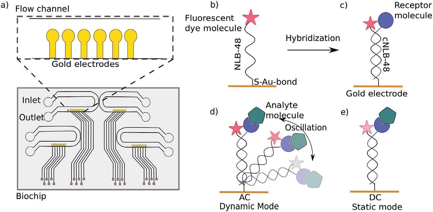
a) Top view of a switchSENSE microfluidic biochip with four flow channels. Each channel comprises six gold electrodes. The more detailed view shows the six independent measurement electrodes in a row within the microfluidic channel. b) Schematic overview of a single stranded DNA nanolever (NL-B48) immobilized on a gold electrode via thiol coupling. The nanolever carries a fluorophore at the lateral end. c) The DNA monolayer is functionalized with the ligand of interest by hybridization of the complementary DNA strand carrying a receptor molecule at the distal end. d) Dynamic measurement mode: The double stranded DNA nanolever is actuated to a switching motion by applying an alternating potential. The fluorescence signal is gradually quenched by energy transfer upon approaching the gold surface. Read-out of this mode is the switching speed of DNA nanolevers. The switching speed is slowed down upon binding of an analyte to the ligand molecule adding friction to the nanolever. e) Static measurement mode: DNA nanolevers are kept at an upright position. Read-out of this mode is the change in fluorescence intensity upon binding of an analyte due to changes in the local environment of the dye. Drawings are not to scale. Image Credit: Kruse M et al., Advanced Material
The ability to calculate not just the dissociation constant but also rate constants is one of the key features of the switchSENSE technique. These kinetic metrics are becoming increasingly important in determining the effectiveness of candidates in medication development.
Other methods for determining dissociation constants exist, but they lack the capacity to resolve bonding kinetic parameters. Radioligand binding tests, microscale thermophoresis (MST), and isothermal calorimetry are some examples of these.
Surface plasmon resonance is one technology that allows for real-time monitoring of kinetic parameters (SPR). SPR measures and switchSENSE technologies are similar in that they both have benefits and drawbacks. As a result, a similar naming system is used.
The quantifiable metrics of kinetic parameters in a timely manner, label-free detection, minimal sample expenditure, and high sensitivity of the technology are all benefits of SPR and switchSENSE. The disadvantages of both procedures are that one of the participants must be immobilized, which may have an impact on binding behavior, and that mass transfer is limited….
News

Our books now available worldwide!
Online Sellers other than Amazon, Routledge, and IOPP Indigo Global Health Care Equivalency in the Age of Nanotechnology, Nanomedicine and Artifcial Intelligence Global Health Care Equivalency In The Age Of Nanotechnology, Nanomedicine And Artificial [...]

Younger Generations Are Aging Faster – and It May Be Fueling a Surge in Cancer
Younger generations may be aging biologically faster than those before them, and that shift could help explain rising rates of cancer at younger ages. For decades, cancer was viewed largely as a disease of [...]

Using Cannabis Could Raise Your Stroke Risk by 37%, Massive Study Reveals
Large-scale evidence suggests cannabis, cocaine, and amphetamines may directly raise stroke risk, including in younger adults. As recreational drug use becomes increasingly common, researchers are uncovering evidence that its health consequences may extend far beyond [...]

Could Vitamin C Be the Secret to Keeping Your Brain Younger?
Lower vitamin C levels were linked to reduced brain volume and weaker neural connectivity in older adults, suggesting a potential connection between nutrition and brain health. Could a common vitamin help preserve the brain [...]

This Deadly Disease Was Wiping Out Humans 5,500 Years Ago
A new study suggests plague was already a deadly threat 5,500 years ago, striking small hunter-gatherer communities long before cities and agriculture emerged. For centuries, plague has been remembered as the disease that devastated [...]

China closing in but US leads in biotech quality, commercial reach, survey finds
SAN DIEGO, June 22 (Reuters) - China, which now conducts more clinical drug trials, opens new tab than the U.S., still lags in the quality and commercial reach of its biomedical science, according to a recent survey, opens new [...]

New method generates renewable supply of progenitor immune cells
In a paper published in Cell, a USC Stem Cell-led team reports a new way of generating a renewable and expandable supply of the progenitor cells that give rise to macrophages. These immune cells help [...]

Scientists Just Discovered a Cellular Survival System That Was Never Supposed To Exist
A surprising backup pathway allows cells to make a crucial amino acid when their primary machinery fails. For decades, biologists believed cells had only one way to access a molecule they cannot live without. New [...]

Artificial cells gain porous membranes, enabling lab reactions and drug release
Artificial cells created in the laboratory offer a wide range of potential applications. Until now, however, their membranes—unlike those of real cells—have been virtually impermeable. Researchers at the Max Planck Institute for Polymer Research, [...]

Popular Weight-Loss Drugs Like Ozempic Linked to Lower Breast Cancer Risk
Ozempic and similar weight-loss drugs were linked to a striking 30% reduction in breast cancer risk in a study of more than 110,000 women. Popular weight-loss and diabetes medications such as Ozempic, Wegovy, Mounjaro, [...]

Stanford Scientists Discover Explosive New Type of Immune Cell
Scientists studying the remarkable regenerative abilities of planarian flatworms have uncovered a previously unknown type of immune cell with an unusually destructive defense strategy. What if an immune cell could wipe out nearby threats [...]

Big Pharma-backed SonoThera sounds off with $125M series B for bubble-based genetic delivery
Bay Area biotech SonoThera is bubbling to a clinical boil after raising a $125 million series B with the backing of some of the biggest names in pharma. Vida Ventures led the raise, with the venture [...]

Joint initiative of 5 EU countries calls for ‘unified approach’ to pharma framework amid US drug pricing pressure
With drug pricing pressure building from the U.S., a healthcare-focused consortium of five European countries is calling for a “unified approach” to strengthen Europe’s pharmaceutical framework and access to innovative medicines. Belgium, the Netherlands, [...]

Molecular Manufacturing: The Future of Nanomedicine – New book from NanoappsMedical Inc.
This book explores the revolutionary potential of atomically precise manufacturing technologies to transform global healthcare, as well as practically every other sector across society. This forward-thinking volume examines how envisaged Factory@Home systems might enable the cost-effective [...]

NanoMedical Brain/Cloud Interface – Explorations and Implications. A new book from Frank Boehm
New book from Frank Boehm, NanoappsMedical Inc Founder: This book explores the future hypothetical possibility that the cerebral cortex of the human brain might be seamlessly, safely, and securely connected with the Cloud via [...]

New book from Nanoappsmedical Inc. – Global Health Care Equivalency
A new book by Frank Boehm, NanoappsMedical Inc. Founder. This groundbreaking volume explores the vision of a Global Health Care Equivalency (GHCE) system powered by artificial intelligence and quantum computing technologies, operating on secure [...]