Fully understanding how the human brain works requires knowing the relationships between the various cells that make up the brain. This entails visualizing the brain’s structure on the scale of nanometers in order to see the connections between neurons.
A team of researchers, led by Dr. Jeff Lichtman at Harvard University and Dr. Viren Jain at Google Research, used electron microscopy (EM) to image a cubic millimeter-sized piece of human brain tissue at high resolution. The tissue was removed from the cerebral cortex of a patient as part of a surgery for epilepsy.
The team began by cutting the tissue into more than 5,000 slices, or sections, each of which was then imaged by EM. This yielded about 1.4 petabytes, or 1,400 terabytes, of data. Using these data, the researchers generated a 3D reconstruction of almost every cell in the sample. Results of the NIH-funded study appeared in Science on May 10, 2024.
Analysis of individual cells in the sample revealed a total of more than 57,000 cells. Most of these were either neurons, which send electrical signals, or glia, which provide various support functions to the neurons. Glia outnumbered neurons 2-to-1. The most common glial cells were oligodendrocytes, which provide structural support and electrical insulation to neurons. The one cubic mm sample also contained about 230 mm of blood vessels.
The reconstruction revealed structural details not seen before. The researchers analyzed a type of neuron, called triangular cells, that are found in the deepest layer of the cerebral cortex. Many of these adopted one of two orientations, which were mirror images of each other. The significance of this organization remains unknown.
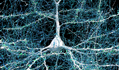
 Researchers built a 3D image of nearly every neuron and its connections within a small piece of human brain tissue.
 The team used machine learning to identify synapses—the junctions through which signals pass from one cell to another. They found almost 150 million synapses. Almost all neurons formed only one synapse with a given target cell. But a small fraction formed two or more synapses to the same target. In at least one case, more than 50 synapses connected a single pair of cells. Although rare, connections of seven or more synapses between cells were much more common than expected by chance. This suggests that these strong connections have some functional significance.
The results illustrate just how complex the brain is at the cellular level. They also show the value of connectomics—the science of generating comprehensive maps of connections between brain cells—for understanding brain function.
“The word ‘fragment’ is ironic,” Lichtman says. “A terabyte is, for most people, gigantic, yet a fragment of a human brain—just a miniscule, teeny-weeny little bit of human brain—is still thousands of terabytes.”
The team has made their dataset available to the public. They have also provided various software tools to help examine the brain map. The hope is that further study of the data, by this team and others, will yield new insight into the workings of the human brain.
“This incredible advancement—the ability to capture and process over 1,000 terabytes of data from the brain—wouldn’t have been possible without a study participant’s generous donation and the important partnerships between neuroscientists, computer scientists, and engineers,” says Dr. John Ngai, director of NIH’s BRAIN Initiative. “These collaborations are central in our aim of building a full map of the human brain so we can bring cures closer to the clinic.”
—by Brian Doctrow, Ph.D.
News

Could Vitamin C Be the Secret to Keeping Your Brain Younger?
Lower vitamin C levels were linked to reduced brain volume and weaker neural connectivity in older adults, suggesting a potential connection between nutrition and brain health. Could a common vitamin help preserve the brain [...]

This Deadly Disease Was Wiping Out Humans 5,500 Years Ago
A new study suggests plague was already a deadly threat 5,500 years ago, striking small hunter-gatherer communities long before cities and agriculture emerged. For centuries, plague has been remembered as the disease that devastated [...]

China closing in but US leads in biotech quality, commercial reach, survey finds
SAN DIEGO, June 22 (Reuters) - China, which now conducts more clinical drug trials, opens new tab than the U.S., still lags in the quality and commercial reach of its biomedical science, according to a recent survey, opens new [...]

New method generates renewable supply of progenitor immune cells
In a paper published in Cell, a USC Stem Cell-led team reports a new way of generating a renewable and expandable supply of the progenitor cells that give rise to macrophages. These immune cells help [...]

Scientists Just Discovered a Cellular Survival System That Was Never Supposed To Exist
A surprising backup pathway allows cells to make a crucial amino acid when their primary machinery fails. For decades, biologists believed cells had only one way to access a molecule they cannot live without. New [...]

Artificial cells gain porous membranes, enabling lab reactions and drug release
Artificial cells created in the laboratory offer a wide range of potential applications. Until now, however, their membranes—unlike those of real cells—have been virtually impermeable. Researchers at the Max Planck Institute for Polymer Research, [...]

Popular Weight-Loss Drugs Like Ozempic Linked to Lower Breast Cancer Risk
Ozempic and similar weight-loss drugs were linked to a striking 30% reduction in breast cancer risk in a study of more than 110,000 women. Popular weight-loss and diabetes medications such as Ozempic, Wegovy, Mounjaro, [...]

Stanford Scientists Discover Explosive New Type of Immune Cell
Scientists studying the remarkable regenerative abilities of planarian flatworms have uncovered a previously unknown type of immune cell with an unusually destructive defense strategy. What if an immune cell could wipe out nearby threats [...]

Big Pharma-backed SonoThera sounds off with $125M series B for bubble-based genetic delivery
Bay Area biotech SonoThera is bubbling to a clinical boil after raising a $125 million series B with the backing of some of the biggest names in pharma. Vida Ventures led the raise, with the venture [...]

Joint initiative of 5 EU countries calls for ‘unified approach’ to pharma framework amid US drug pricing pressure
With drug pricing pressure building from the U.S., a healthcare-focused consortium of five European countries is calling for a “unified approach” to strengthen Europe’s pharmaceutical framework and access to innovative medicines. Belgium, the Netherlands, [...]

Our books now available worldwide!
Online Sellers other than Amazon, Routledge, and IOPP Indigo Global Health Care Equivalency in the Age of Nanotechnology, Nanomedicine and Artifcial Intelligence Global Health Care Equivalency In The Age Of Nanotechnology, Nanomedicine And Artificial [...]

Molecular Manufacturing: The Future of Nanomedicine – New book from NanoappsMedical Inc.
This book explores the revolutionary potential of atomically precise manufacturing technologies to transform global healthcare, as well as practically every other sector across society. This forward-thinking volume examines how envisaged Factory@Home systems might enable the cost-effective [...]

NanoMedical Brain/Cloud Interface – Explorations and Implications. A new book from Frank Boehm
New book from Frank Boehm, NanoappsMedical Inc Founder: This book explores the future hypothetical possibility that the cerebral cortex of the human brain might be seamlessly, safely, and securely connected with the Cloud via [...]

New book from Nanoappsmedical Inc. – Global Health Care Equivalency
A new book by Frank Boehm, NanoappsMedical Inc. Founder. This groundbreaking volume explores the vision of a Global Health Care Equivalency (GHCE) system powered by artificial intelligence and quantum computing technologies, operating on secure [...]
UCLA Scientists Uncover a “Hidden Weakness” in Some of the World’s Deadliest Cancers
A new study has uncovered an unexpected vulnerability in some of the deadliest cancers. Researchers at UCLA have identified a previously hidden weakness in some of the most aggressive cancers, pointing to a possible new way [...]
AI-designed universal coronavirus vaccine clears first human trial
Key Takeaways Super-Antigen Technology: Uses AI and machine learning to analyze viral genomes, creating a single vaccine that targets essential features across entire virus families, including coronaviruses and Ebola. Human Trials & Safety: Phase [...]