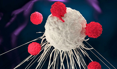
Exciting and novel research has investigated the internalization of nanoparticles by cells to optimize drug delivery into target cells. This research, published within the journal, Pharmaceutics, aims to analyze the simultaneous uptake of two different types of nanoparticles into cells.
Enhancing Drug Delivery Using Nanoparticles
Nanoparticles, sized within the nanoscale, between 1 and 100 nm, hold high efficacy for drug delivery due to their ability to interact with biological systems.
The use of nanocarriers to carry drugs to their target cells is one of the advantages of incorporating nanotechnology within medicine, especially with their surface functionalization ability which enables them to target cells more effectively. This allows them to be more beneficial than the conventional method of drug delivery which has a systemic effect and can cause adverse effects on all cells rather than just targeting specific cells.
Understanding how many carriers enter cells is critical for deciphering the intercellular dose that the target cells are provided to determine the efficacy of utilizing nanoparticles in this way and advancing drug delivery systems.
While this area of research has already been spearheaded with previous research with internalization pathways for particles being investigated, the effect of cell uptake when two different particles are exposed to the cell simultaneously is limited.
These investigations include reports of an increase in cell uptake of gold and iron nanoparticles when both particles were exposed to murine macrophages. However, another researcher has described changes to cellular function with simultaneous exposure, with carbon black and iron oxide particles exposed to human epithelial cells, possibly resulting in protein and lipid oxidation.
Interestingly, this novel research can be useful when enhancing drug delivery in the body by using two different particles that can be used for various applications to increase the efficacy of drug treatment for diseases and disorders.
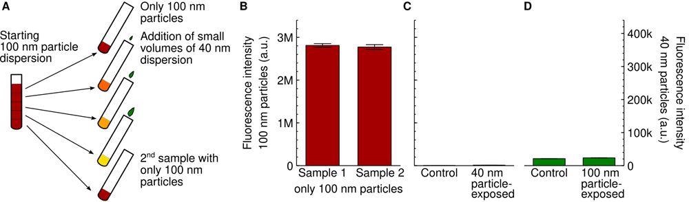
Experimental setup. (A) Schematic showing the dispersion procedure we used to ensure that the concentration of the 100 nm particles was the same for all samples. A first dispersion of the 100 nm particles was prepared and then divided up. To the resulting samples, different small volumes of a 40 nm particle dispersion were added. The first and last sample were left without 40 nm particles to serve as controls. Note that the colours are schematic only. (B) Nanoparticle fluorescence of cells exposed to the two different control dispersions of the 100 nm particles mentioned in panel A (first and last sample; 20 μg/mL or 0.060 nM). The similarity of the signal is consistent with the two control samples having the same nanoparticle concentration. (C,D) Lack of cross-talk between the two particle signals. (C) Cells were exposed to only the 40 nm particles (100 μg/mL; 4.7 nM) and the fluorescence intensity measured at the same wavelengths where researchers measured the 100 nm particles (left axis). There is a low background signal (right bar), but this is comparable to control cells not exposed to any particles at all (left bar). (D) Cells were exposed to only the 100 nm particles (80 μg/mL; 0.24 nM) and the fluorescence intensity measured at the same wavelengths where we measure the 40 nm particles (right axis). There is a low background signal (right bar), but this is comparable to control cells not exposed to any particles at all (left bar). © De Boer I, Richards CJ, Åberg C. (2022)
Innovative Research
The researchers of this study exposed HeLa (adenocarcinomic human cervical epithelial) cells to two different sized carboxylated polystyrene nanoparticles, including 40 nm and 100 nm diameter particles labeled with different fluorescent dyes to track the movement.
They observed that smaller particles had an increased cell uptake in the presence of larger-sized particles while larger particle uptake was impeded in the presence of smaller particles. This confirmed previous research on simultaneous cell uptake.
However, this increase was confirmed of cells within a medium with serum and the formation of a biomolecular corona on particles, which was not the same as previous research within a serum-free medium.
This research can enable the understanding of whether the competition between different concentrations of various sized particles is dependent on cell types; through the examination of 2D fluorescence distribution of the two measurable particles, the results have illustrated all cells take up both particles, but this may be within varying levels.
A limitation of this research is a lack of comprehension about the uptake mechanisms underlying the competitive nature of two various sized nanoparticles. With further research, the cellular mechanisms of this uptake can be clarified to understand these observations.
Significant Implications for Enhanced Drug Delivery
This research can be utilized to enhance drug delivery with the presence of a second nanoparticle, such as one of a larger size with the aim of increasing the cellular uptake of the first nanoparticle. The implications of this can be significant for increasing the intercellular drug dose found within cells and can be a start to advancing nanotechnology-based drug delivery systems.
With nanoparticles already being able to be functionalized through surface ligands, the use of these as nanocarriers for drug delivery as well as secondary particles can be an innovative approach to increasing the therapeutic selectivity of cells.
This can be utilized for diseases and even for cancer treatment to develop particles that work together for enhanced drug delivery into target cells, sustained delivery, and possibly the clearance of the drug from the body. Innovative drug delivery systems with more control through this approach may be a step towards personalized medicine that prioritizes patient care.
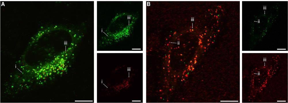
Subcellular distribution of the 40 nm and 100 nm particles. Cells were exposed for 24 h to both the 40 nm and 100 nm particles simultaneously and then observed using confocal fluorescence microscopy. (A) Concentration of 100 and 40 nm particles 20 and 100 μg/mL, respectively (0.060 nM and 4.7 nM; conditions correspond to the highest 40 nm particle concentration in Figures 2 and S3). (B) Concentration of 40 and 100 nm particles 6.25 and 80 μg/mL, respectively (0.30 and 0.24 nM; conditions correspond to the highest 100 nm particle concentration in Figures 4 and S4). The larger images show overlaps of both fluorescence colours, while the smaller images show the individual colours. (Green) 40 nm particles; (red) 100 nm particles. Arrows show examples of (i) 40 nm particle( s) (green) in the absence of 100 nm particles; (ii) 100 nm particle(s) (red) in the absence of 40 nm particles; (iii) 40 and 100 nm particles in the same location. The results show that the two particles often end up in the same location, but not always. All scale bars correspond to 10 μm. © De Boer I, Richards CJ, Åberg C. (2022)
News

China closing in but US leads in biotech quality, commercial reach, survey finds
SAN DIEGO, June 22 (Reuters) - China, which now conducts more clinical drug trials, opens new tab than the U.S., still lags in the quality and commercial reach of its biomedical science, according to a recent survey, opens new [...]

New method generates renewable supply of progenitor immune cells
In a paper published in Cell, a USC Stem Cell-led team reports a new way of generating a renewable and expandable supply of the progenitor cells that give rise to macrophages. These immune cells help [...]

Scientists Just Discovered a Cellular Survival System That Was Never Supposed To Exist
A surprising backup pathway allows cells to make a crucial amino acid when their primary machinery fails. For decades, biologists believed cells had only one way to access a molecule they cannot live without. New [...]

Artificial cells gain porous membranes, enabling lab reactions and drug release
Artificial cells created in the laboratory offer a wide range of potential applications. Until now, however, their membranes—unlike those of real cells—have been virtually impermeable. Researchers at the Max Planck Institute for Polymer Research, [...]

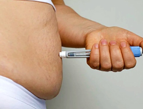
Popular Weight-Loss Drugs Like Ozempic Linked to Lower Breast Cancer Risk
Ozempic and similar weight-loss drugs were linked to a striking 30% reduction in breast cancer risk in a study of more than 110,000 women. Popular weight-loss and diabetes medications such as Ozempic, Wegovy, Mounjaro, [...]

Stanford Scientists Discover Explosive New Type of Immune Cell
Scientists studying the remarkable regenerative abilities of planarian flatworms have uncovered a previously unknown type of immune cell with an unusually destructive defense strategy. What if an immune cell could wipe out nearby threats [...]

Big Pharma-backed SonoThera sounds off with $125M series B for bubble-based genetic delivery
Bay Area biotech SonoThera is bubbling to a clinical boil after raising a $125 million series B with the backing of some of the biggest names in pharma. Vida Ventures led the raise, with the venture [...]

Joint initiative of 5 EU countries calls for ‘unified approach’ to pharma framework amid US drug pricing pressure
With drug pricing pressure building from the U.S., a healthcare-focused consortium of five European countries is calling for a “unified approach” to strengthen Europe’s pharmaceutical framework and access to innovative medicines. Belgium, the Netherlands, [...]

Our books now available worldwide!
Online Sellers other than Amazon, Routledge, and IOPP Indigo Global Health Care Equivalency in the Age of Nanotechnology, Nanomedicine and Artifcial Intelligence Global Health Care Equivalency In The Age Of Nanotechnology, Nanomedicine And Artificial [...]

Molecular Manufacturing: The Future of Nanomedicine – New book from NanoappsMedical Inc.
This book explores the revolutionary potential of atomically precise manufacturing technologies to transform global healthcare, as well as practically every other sector across society. This forward-thinking volume examines how envisaged Factory@Home systems might enable the cost-effective [...]

NanoMedical Brain/Cloud Interface – Explorations and Implications. A new book from Frank Boehm
New book from Frank Boehm, NanoappsMedical Inc Founder: This book explores the future hypothetical possibility that the cerebral cortex of the human brain might be seamlessly, safely, and securely connected with the Cloud via [...]

New book from Nanoappsmedical Inc. – Global Health Care Equivalency
A new book by Frank Boehm, NanoappsMedical Inc. Founder. This groundbreaking volume explores the vision of a Global Health Care Equivalency (GHCE) system powered by artificial intelligence and quantum computing technologies, operating on secure [...]
UCLA Scientists Uncover a “Hidden Weakness” in Some of the World’s Deadliest Cancers
A new study has uncovered an unexpected vulnerability in some of the deadliest cancers. Researchers at UCLA have identified a previously hidden weakness in some of the most aggressive cancers, pointing to a possible new way [...]
AI-designed universal coronavirus vaccine clears first human trial
Key Takeaways Super-Antigen Technology: Uses AI and machine learning to analyze viral genomes, creating a single vaccine that targets essential features across entire virus families, including coronaviruses and Ebola. Human Trials & Safety: Phase [...]

Researchers Discover a Hidden Vitamin D Problem That Persists Year-Round
A new study suggests that some groups may not experience the expected seasonal boost in vitamin D levels, even during the sunniest months of the year. Many people assume that spending more time outdoors [...]
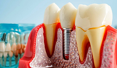
Researchers Solve the Mystery Behind a Billion-Dollar Dental Implant Disease
Researchers have uncovered why a common and costly dental implant infection often resists antibiotics. Dental implants have helped tens of millions of people regain a full set of stable, functional teeth, something traditional dentures [...]