Artificial intelligence facilitates the visualization of neural connections in the brains of mice.
Scientists from Johns Hopkins have leveraged artificial intelligence to create a technique that allows for the visualization and monitoring of alterations in the strength of synapses — the connection points through which nerve cells in the brain communicate — in living organisms. The technique, as outlined in Nature Methods, could, according to the researchers, pave the way for an improved comprehension of how these connections in human brains evolve with learning, age, trauma, and disease.
"If you want to learn more about how an orchestra plays, you have to watch individual players over time, and this new method does that for synapses in the brains of living animals," says Dwight Bergles, Ph.D., the Diana Sylvestre and Charles Homcy Professor in the Solomon H. Snyder Department of Neuroscience at the Johns Hopkins University (JHU) School of Medicine.
Bergles co-authored the study with colleagues Adam Charles, Ph.D., M.E., and Jeremias Sulam, Ph.D., both assistant professors in the biomedical engineering department, and Richard Huganir, Ph.D., Bloomberg Distinguished Professor at JHU and Director of the Solomon H. Snyder Department of Neuroscience. All four researchers are members of Johns Hopkins' Kavli Neuroscience Discovery Institute.
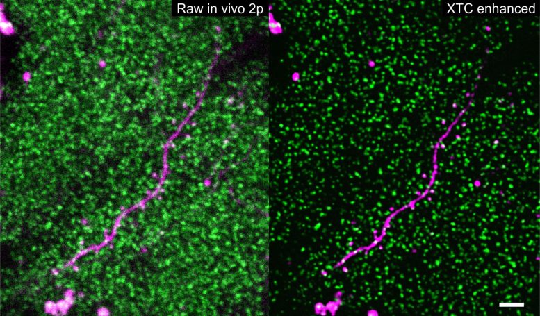
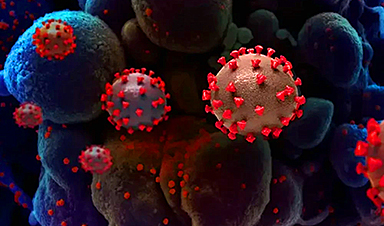
Thousands of SEP-GluA2 tagged synapses (green) surrounding a sparsely labeled dendrite (magenta) before and after XTC image resolution enhancement. Scale bar 5 microns. Credit: Xu, Y.K.T., Graves, A.R., Coste, G.I. et al. Nat Methods
Nerve cells transfer information from one cell to another by exchanging chemical messages at synapses ("junctions"). In the brain, the authors explain, different life experiences, such as exposure to new environments and learning skills, are thought to induce changes at synapses, strengthening or weakening these connections to allow learning and memory. Understanding how these minute changes occur across the trillions of synapses in our brains is a daunting challenge, but it is central to uncovering how the brain works when healthy and how it is altered by disease.
To determine which synapses change during a particular life event, scientists have long sought better ways to visualize the shifting chemistry of synaptic messaging, necessitated by the high density of synapses in the brain and their small size — traits that make them extremely hard to visualize even with new state-of-the-art microscopes.
"We needed to go from challenging, blurry, noisy imaging data to extract the signal portions we need to see," Charles says.
To do so, Bergles, Sulam, Charles, Huganir, and their colleagues turned to machine learning, a computational framework that allows the flexible development of automatic data processing tools. Machine learning has been successfully applied to many domains across biomedical imaging, and in this case, the scientists leveraged the approach to enhance the quality of images composed of thousands of synapses. Although it can be a powerful tool for automated detection, greatly surpassing human speeds, the system must first be "trained," teaching the algorithm what high-quality images of synapses should look like.
In these experiments, the researchers worked with genetically altered mice in which glutamate receptors — the chemical sensors at synapses — glowed green (fluoresced) when exposed to light. Because each receptor emits the same amount of light, the amount of fluorescence generated by a synapse in these mice is an indication of the number of synapses, and therefore its strength.
As expected, imaging in the intact brain produced low-quality pictures in which individual clusters of glutamate receptors at synapses were difficult to see clearly, let alone to be individually detected and tracked over time. To convert these into higher-quality images, the scientists trained a machine learning algorithm with images taken of brain slices (ex vivo) derived from the same type of genetically altered mice. Because these images weren't from living animals, it was possible to produce much higher quality images using a different microscopy technique, as well as low-quality images — similar to those taken in live animals — of the same views.
This cross-modality data collection framework enabled the team to develop an enhancement algorithm that can produce higher-resolution images from low-quality ones, similar to the images collected from living mice. In this way, data collected from the intact brain can be significantly enhanced and able to detect and track individual synapses (in the thousands) during multiday experiments.
To follow changes in receptors over time in living mice, the researchers then used microscopy to take repeated images of the same synapses in mice over several weeks. After capturing baseline images, the team placed the animals in a chamber with new sights, smells, and tactile stimulation for a single five-minute period. They then imaged the same area of the brain every other day to see if and how the new stimuli had affected the number of glutamate receptors at synapses.
Although the focus of the work was on developing a set of methods to analyze synapse level changes in many different contexts, the researchers found that this simple change in environment caused a spectrum of alterations in fluorescence across synapses in the cerebral cortex, indicating connections where the strength increased and others where it decreased, with a bias toward strengthening in animals exposed to the novel environment.
The studies were enabled through close collaboration among scientists with distinct expertise, ranging from molecular biology to artificial intelligence, who don't normally work closely together. But such collaboration, is encouraged at the cross-disciplinary Kavli Neuroscience Discovery Institute, Bergles says. The researchers are now using this machine learning approach to study synaptic changes in animal models of Alzheimer's disease, and they believe the method could shed new light on synaptic changes that occur in other disease and injury contexts.
"We are really excited to see how and where the rest of the scientific community will take this," Sulam says.
Reference: "Cross-modality supervised image restoration enables nanoscale tracking of synaptic plasticity in living mice" by Yu Kang T. Xu, Austin R. Graves, Gabrielle I. Coste, Richard L. Huganir, Dwight E. Bergles, Adam S. Charles and Jeremias Sulam, 11 May 2023, Nature Methods.
DOI: 10.1038/s41592-023-01871-6
News

This Deadly Disease Was Wiping Out Humans 5,500 Years Ago
A new study suggests plague was already a deadly threat 5,500 years ago, striking small hunter-gatherer communities long before cities and agriculture emerged. For centuries, plague has been remembered as the disease that devastated [...]

China closing in but US leads in biotech quality, commercial reach, survey finds
SAN DIEGO, June 22 (Reuters) - China, which now conducts more clinical drug trials, opens new tab than the U.S., still lags in the quality and commercial reach of its biomedical science, according to a recent survey, opens new [...]

New method generates renewable supply of progenitor immune cells
In a paper published in Cell, a USC Stem Cell-led team reports a new way of generating a renewable and expandable supply of the progenitor cells that give rise to macrophages. These immune cells help [...]

Scientists Just Discovered a Cellular Survival System That Was Never Supposed To Exist
A surprising backup pathway allows cells to make a crucial amino acid when their primary machinery fails. For decades, biologists believed cells had only one way to access a molecule they cannot live without. New [...]

Artificial cells gain porous membranes, enabling lab reactions and drug release
Artificial cells created in the laboratory offer a wide range of potential applications. Until now, however, their membranes—unlike those of real cells—have been virtually impermeable. Researchers at the Max Planck Institute for Polymer Research, [...]

Popular Weight-Loss Drugs Like Ozempic Linked to Lower Breast Cancer Risk
Ozempic and similar weight-loss drugs were linked to a striking 30% reduction in breast cancer risk in a study of more than 110,000 women. Popular weight-loss and diabetes medications such as Ozempic, Wegovy, Mounjaro, [...]

Stanford Scientists Discover Explosive New Type of Immune Cell
Scientists studying the remarkable regenerative abilities of planarian flatworms have uncovered a previously unknown type of immune cell with an unusually destructive defense strategy. What if an immune cell could wipe out nearby threats [...]

Big Pharma-backed SonoThera sounds off with $125M series B for bubble-based genetic delivery
Bay Area biotech SonoThera is bubbling to a clinical boil after raising a $125 million series B with the backing of some of the biggest names in pharma. Vida Ventures led the raise, with the venture [...]

Joint initiative of 5 EU countries calls for ‘unified approach’ to pharma framework amid US drug pricing pressure
With drug pricing pressure building from the U.S., a healthcare-focused consortium of five European countries is calling for a “unified approach” to strengthen Europe’s pharmaceutical framework and access to innovative medicines. Belgium, the Netherlands, [...]

Our books now available worldwide!
Online Sellers other than Amazon, Routledge, and IOPP Indigo Global Health Care Equivalency in the Age of Nanotechnology, Nanomedicine and Artifcial Intelligence Global Health Care Equivalency In The Age Of Nanotechnology, Nanomedicine And Artificial [...]

Molecular Manufacturing: The Future of Nanomedicine – New book from NanoappsMedical Inc.
This book explores the revolutionary potential of atomically precise manufacturing technologies to transform global healthcare, as well as practically every other sector across society. This forward-thinking volume examines how envisaged Factory@Home systems might enable the cost-effective [...]

NanoMedical Brain/Cloud Interface – Explorations and Implications. A new book from Frank Boehm
New book from Frank Boehm, NanoappsMedical Inc Founder: This book explores the future hypothetical possibility that the cerebral cortex of the human brain might be seamlessly, safely, and securely connected with the Cloud via [...]

New book from Nanoappsmedical Inc. – Global Health Care Equivalency
A new book by Frank Boehm, NanoappsMedical Inc. Founder. This groundbreaking volume explores the vision of a Global Health Care Equivalency (GHCE) system powered by artificial intelligence and quantum computing technologies, operating on secure [...]
UCLA Scientists Uncover a “Hidden Weakness” in Some of the World’s Deadliest Cancers
A new study has uncovered an unexpected vulnerability in some of the deadliest cancers. Researchers at UCLA have identified a previously hidden weakness in some of the most aggressive cancers, pointing to a possible new way [...]
AI-designed universal coronavirus vaccine clears first human trial
Key Takeaways Super-Antigen Technology: Uses AI and machine learning to analyze viral genomes, creating a single vaccine that targets essential features across entire virus families, including coronaviruses and Ebola. Human Trials & Safety: Phase [...]

Researchers Discover a Hidden Vitamin D Problem That Persists Year-Round
A new study suggests that some groups may not experience the expected seasonal boost in vitamin D levels, even during the sunniest months of the year. Many people assume that spending more time outdoors [...]