A team of researchers recently published a paper in the journal ACS Applied Nano Materials that demonstrated the effectiveness of specific chemically-modified nanohole edges in reduced graphene oxide (rGO) in binding proteins.
Graphene-Based Biosensors
Biosensors are gaining considerable prominence owing to the rising significance of quantitative or qualitative detection of biological markers and early prognosis of diseases. Such a shift is necessitating the development of accessible, accurate, and rapid techniques.
Biosensors based on graphene, where graphene is used as a transducer or buffer, have attracted wide attention due to the exceptional properties of graphene, such as effect-free high permittivity and high carrier mobility.
Graphene combined with nanoparticles has been investigated as a biosensor in previous research; however, during the development of such biosensors, certain properties of graphene can potentially affect the detection limit of the sensor. Thus, the surface specificity of graphene must be modified for certain molecules to use in biosensors.
Modifying Graphene Nanohole Edges
In this study, researchers initially synthesized rGO nanomembrane (NMG), with nanoholes in the scale of 40-60 nm center-to-center distances and 20-25 nm in diameter, following two distinct approaches that are based on gold nano-islands (Au-NIs) and gold nanoparticles (AuNPs). Later, they performed a specific covalent chemical modification of the nanohole edges in the NMG and evaluated their effectiveness in capturing biological molecules.
rGO-AuNPs were fabricated by depositing an rGO monolayer over a self-assembled AuNP monolayer and then removing the NPs. rGO-Au-NIs were also formed in a similar manner excluding the self-assembled AuNP monolayer, which was replaced by an Au-NI layer.
Ultraviolet (UV) spectroscopy, infrared spectroscopy, and scanning electron microscope (SEM) were used to characterize the deposited layers after and before removing the NIs or the NPs.
The chemical groups in the layers were defined and investigated by Fourier transform infrared (FTIR) spectroscopy.
3-(aminopropyl) triethoxysilane (APTES) was utilized to modify the holes’ edges, while N-ethyl-N′-(3-(dimethylamino) propyl) carbodiimide/N-hydroxysuccinimide) (EDC/NHS) was applied as coupling agents to facilitate coupling between the NMG nanohole edges and a biological moiety such as an antibody.
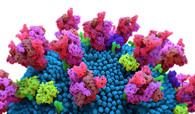
The successful modification of the NMG edges and the scope of monitoring protein binding were investigated by attaching the angiotensin-converting enzyme 2 (ACE2) antibody to the edges, and subsequently measuring the binding ability of the edges to severe acute respiratory syndrome coronavirus 2 (SARS-CoV-2) spike protein.
A number of control experiments were performed to verify the successful specific binding between the antibody/bioreceptor/ACE2 protein and the target molecule/SARS-CoV-2 spike protein.
Achieving Successful Protein Binding
NMG was successfully synthesized on the rGO layer using AuNPs and Au-NIs. The rGO films appeared as separated wrinkled flakes owing to the deformation after exfoliation and restacking processes, indicating the formation of a high-quality rGO-layered structure nanosheet.
The flakes appeared as a uniformed background after the rGO monolayer was deposited on the glass slide, and demonstrated a 97.7% transparency at a wavelength of 550 nm.
The FTIR spectrum of the GO layer displayed characteristic peaks of hydroxyl, carboxyl, aromatic, carboxy, epoxy group, whereas the FTIR spectrum of the chemically reduced GO or rGO lacked the peaks of oxygen groups that include epoxy and hydroxyl groups. Additionally, the intensities of characteristic peaks of oxygen were reduced in rGO, indicating a high degree of reduction of the GO sheet.
The aromatic groups represented the only remaining peaks at 1585 and 1623 cm-1 in the rGO.
NIs were distributed homogenously with a diameter of 25-35 nm after annealing. A high concentration of hydroxyl groups was observed at the nanohole edges in the NMG after its formation. The absorption peaks associated with nitrogen-hydrogen (N−H) bending and carbon-hydrogen (C−H) swing were observed at 1582 and 1380 cm-1 after the NMG was silanized with APTES, indicating the functionalization of NMG with APTES.
After the functionalization, the amine group was observed in the NMG with a separate peak at 1414 cm-1, which corresponded to the oxygen-hydrogen (O−H) bending vibration of the carboxyl group.
An amide bond was observed at 1533 cm-1 after the application of coupling agents at NMG edges, and more bands at 3280 and 1641 cm-1 emerged after the covalent bond interactions with the ACE2.
The amide bond was created between the carboxyl groups on the antibody and APTES-modified NMG edges owing to the presence of the amine groups at the edges. The intensity of the 1641 cm-1 peak after antibody interaction was higher compared to the peak observed in the NH2-NMG, indicating the successful antibody surface functionalization on NH2-NMG.
The FTIR spectra of NMG and rGO were measured by modifying and activating both of them with APTES and EDC/NHS. No change in peak positions was observed in rGO. However, the intensity of the amide bond increased substantially in NMG, indicating the specificity of protein binding only to hole edges and not to the surface.
Real-time binding measurements demonstrated an affinity constant of 0.93 × 10⁹ M-1 and a dissociation constant (KD) of 1.08 nM.
Taken together, the findings of this study demonstrated a versatile and robust mechanism to perform specific modifications of NMG edges to use NMG as a real-time highly sensitive biosensor.
News

Our books now available worldwide!
Online Sellers other than Amazon, Routledge, and IOPP Indigo Global Health Care Equivalency in the Age of Nanotechnology, Nanomedicine and Artifcial Intelligence Global Health Care Equivalency In The Age Of Nanotechnology, Nanomedicine And Artificial [...]

Younger Generations Are Aging Faster – and It May Be Fueling a Surge in Cancer
Younger generations may be aging biologically faster than those before them, and that shift could help explain rising rates of cancer at younger ages. For decades, cancer was viewed largely as a disease of [...]

Using Cannabis Could Raise Your Stroke Risk by 37%, Massive Study Reveals
Large-scale evidence suggests cannabis, cocaine, and amphetamines may directly raise stroke risk, including in younger adults. As recreational drug use becomes increasingly common, researchers are uncovering evidence that its health consequences may extend far beyond [...]

Could Vitamin C Be the Secret to Keeping Your Brain Younger?
Lower vitamin C levels were linked to reduced brain volume and weaker neural connectivity in older adults, suggesting a potential connection between nutrition and brain health. Could a common vitamin help preserve the brain [...]

This Deadly Disease Was Wiping Out Humans 5,500 Years Ago
A new study suggests plague was already a deadly threat 5,500 years ago, striking small hunter-gatherer communities long before cities and agriculture emerged. For centuries, plague has been remembered as the disease that devastated [...]

China closing in but US leads in biotech quality, commercial reach, survey finds
SAN DIEGO, June 22 (Reuters) - China, which now conducts more clinical drug trials, opens new tab than the U.S., still lags in the quality and commercial reach of its biomedical science, according to a recent survey, opens new [...]

New method generates renewable supply of progenitor immune cells
In a paper published in Cell, a USC Stem Cell-led team reports a new way of generating a renewable and expandable supply of the progenitor cells that give rise to macrophages. These immune cells help [...]

Scientists Just Discovered a Cellular Survival System That Was Never Supposed To Exist
A surprising backup pathway allows cells to make a crucial amino acid when their primary machinery fails. For decades, biologists believed cells had only one way to access a molecule they cannot live without. New [...]

Artificial cells gain porous membranes, enabling lab reactions and drug release
Artificial cells created in the laboratory offer a wide range of potential applications. Until now, however, their membranes—unlike those of real cells—have been virtually impermeable. Researchers at the Max Planck Institute for Polymer Research, [...]

Popular Weight-Loss Drugs Like Ozempic Linked to Lower Breast Cancer Risk
Ozempic and similar weight-loss drugs were linked to a striking 30% reduction in breast cancer risk in a study of more than 110,000 women. Popular weight-loss and diabetes medications such as Ozempic, Wegovy, Mounjaro, [...]

Stanford Scientists Discover Explosive New Type of Immune Cell
Scientists studying the remarkable regenerative abilities of planarian flatworms have uncovered a previously unknown type of immune cell with an unusually destructive defense strategy. What if an immune cell could wipe out nearby threats [...]

Big Pharma-backed SonoThera sounds off with $125M series B for bubble-based genetic delivery
Bay Area biotech SonoThera is bubbling to a clinical boil after raising a $125 million series B with the backing of some of the biggest names in pharma. Vida Ventures led the raise, with the venture [...]

Joint initiative of 5 EU countries calls for ‘unified approach’ to pharma framework amid US drug pricing pressure
With drug pricing pressure building from the U.S., a healthcare-focused consortium of five European countries is calling for a “unified approach” to strengthen Europe’s pharmaceutical framework and access to innovative medicines. Belgium, the Netherlands, [...]

Molecular Manufacturing: The Future of Nanomedicine – New book from NanoappsMedical Inc.
This book explores the revolutionary potential of atomically precise manufacturing technologies to transform global healthcare, as well as practically every other sector across society. This forward-thinking volume examines how envisaged Factory@Home systems might enable the cost-effective [...]

NanoMedical Brain/Cloud Interface – Explorations and Implications. A new book from Frank Boehm
New book from Frank Boehm, NanoappsMedical Inc Founder: This book explores the future hypothetical possibility that the cerebral cortex of the human brain might be seamlessly, safely, and securely connected with the Cloud via [...]

New book from Nanoappsmedical Inc. – Global Health Care Equivalency
A new book by Frank Boehm, NanoappsMedical Inc. Founder. This groundbreaking volume explores the vision of a Global Health Care Equivalency (GHCE) system powered by artificial intelligence and quantum computing technologies, operating on secure [...]