What if you could take a picture of every gene inside a living organism—not with light, but with DNA itself?
Scientists at the University of Chicago have pioneered a revolutionary imaging technique called volumetric DNA microscopy. It builds intricate 3D maps of genetic material by tagging and tracking molecular interactions, creating never-before-seen views inside organisms like zebrafish embryos.
New Window into Genetics
Traditional genetic sequencing can reveal a lot about the genetic material in a sample, such as a piece of tissue or a drop of blood, but it doesn’t show where specific genetic sequences are located within that sample, or how they relate to nearby genes and molecules.
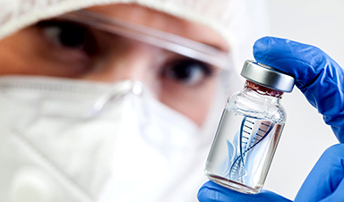
To address this, researchers at the University of Chicago are developing a new technology that captures both the identity and location of genetic material. The method works by tagging individual DNA or RNA molecules and tracking how neighboring tags interact. These interactions are used to build a molecular network that reflects the spatial arrangement of genes, effectively creating a three-dimensional map of genetic activity. Known as volumetric DNA microscopy, the technique generates detailed 3D images of entire organisms from the inside out – down to the level of individual cells.

Imaging an Entire Organism
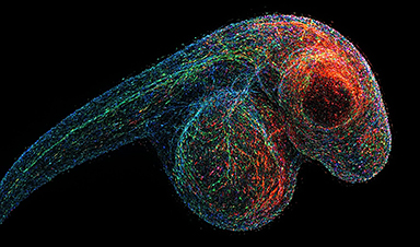
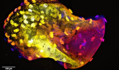
Joshua Weinstein, PhD, Assistant Professor of Medicine and Molecular Engineering at UChicago, has spent over a decade developing DNA microscopy, with support from the National Institutes of Health and the National Science Foundation. In a recent study published today (March 27) in Nature Biotechnology, Weinstein and postdoctoral researcher Nianchao Qian used the technique to produce a complete 3D DNA map of a zebrafish embryo—a widely used model for studying development and the nervous system.
“It’s a level of biology that no one has ever seen before,” Weinstein said. “To be able to see that kind of a view of nature from within a specimen is exhilarating.”
Rethinking Microscopy
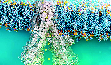
Unlike traditional microscopes that use light or lenses, DNA microscopy creates images by calculating interactions among molecules, providing a new way to visualize genetic material in 3D. First, short DNA sequence tags called unique molecular identifiers (UMIs) are added to cells. They attach to DNA and RNA molecules and begin making copies of themselves. This starts a chemical reaction that creates new sequences, called unique event identifiers (UEIs), that are unique to each pairing.
It’s these pairings that help create the spatial map of where each genetic molecule is located. UMI pairs that are close together interact more frequently and generate more UEIs than those that are farther apart. Once the DNA and RNA are sequenced, a computational model reconstructs their original locations by analyzing the physical links between UMI-tags, creating a spatial map of gene expression.
Cell Phones and Cells: A Clever Analogy
Weinstein compares the technique to using data from cell phones pinging each other to determine people’s location in a city. Knowing the cell phone number or IP address of each person is like knowing the genetic sequence of one molecule, but if you can layer on their interactions with other phones nearby, you can work out their locations too.
“We can do this with cell phones and people, so why not do that with molecules and cells,” he said. “This turns the idea of imaging on its head. Rather than relying on an optical apparatus to shine light in, we can use biochemistry and DNA to form a massive network between molecules and encode their proximities to each other.”
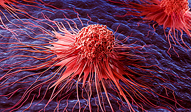
Future Applications in Cancer and Immunotherapy
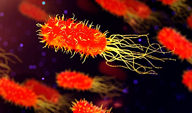
DNA microscopy doesn’t rely on prior knowledge of the genome or shape of a specimen, so it could be useful for understanding genetic expression in unique, unknown contexts. Tumors generate countless new genetic mutations, for example, so the tool would be able to map out the tumor microenvironment and where it interacts with the immune system. Immune cells interact with each other and respond to pathogens in context-specific ways, so DNA microscopy could help unravel those genetic mechanisms. Such applications could in turn guide more precise immunotherapy for cancer or tailor personalized vaccines.
“This is the critical foundation for being able to have truly comprehensive information about the ensemble of unique cells within the lymphatic system or tumor tissue,” Weinstein said. “There has still been this major gap in technology for allowing us to understand idiosyncratic tissue, and that’s what we’re trying to fill in here.”
DOI: 27 March 2025, Nature Biotechnology.
10.1038/s41587-025-02613-z
Additional funding for the study, “Spatial-transcriptomic imaging of an intact organism using volumetric DNA microscopy,” was provided by the Damon Runyon Foundation and the Moore Foundation.
News
Scientists Uncover Fatal Weakness in “Zombie Cells” Linked to Cancer
A newly identified weakness in “zombie” cells may open the door to more precise cancer treatments by turning their own survival strategy against them. A new class of drugs takes advantage of a recently [...]

Bowel and Ovarian Cancers Are Dramatically Rising in Young Adults, Scientists Aren’t Sure Why
Cancer incidence is increasing, especially among younger adults, and current risk factors don’t fully account for the trend. Scientists suggest other underlying causes may be contributing. Cancer patterns in England are shifting in a [...]
New Immune Pathway Could Supercharge mRNA Cancer Vaccines
A surprising backup system in the immune response to mRNA vaccines may hold the key to more effective cancer treatments. The arrival of mRNA vaccines against SARS-CoV-2 in 2020 marked a turning point in the COVID-19 pandemic. Today, [...]
Scientists Discover “Molecular Switch” That Fuels Alzheimer’s Brain Inflammation
A newly identified trigger of brain inflammation could offer a fresh target for slowing Alzheimer’s progression. The brain has its own built-in immune system that identifies threats and responds to them. In Alzheimer’s disease, growing evidence [...]

Molecular Manufacturing: The Future of Nanomedicine – New book from NanoappsMedical Inc.
This book explores the revolutionary potential of atomically precise manufacturing technologies to transform global healthcare, as well as practically every other sector across society. This forward-thinking volume examines how envisaged Factory@Home systems might enable the cost-effective [...]
Forgotten Medicinal Plant Shows Promise in Fighting Dangerous Superbugs
A traditional medicinal plant, tormentil, shows promise against antibiotic-resistant bacteria in laboratory tests. Its compounds work by limiting bacterial growth and boosting antibiotic performance. Before the development of modern antibiotics, plant-based remedies were commonly [...]

NanoMedical Brain/Cloud Interface – Explorations and Implications. A new book from Frank Boehm
New book from Frank Boehm, NanoappsMedical Inc Founder: This book explores the future hypothetical possibility that the cerebral cortex of the human brain might be seamlessly, safely, and securely connected with the Cloud via [...]

New Research Finds Shocking Link Between Chili Peppers and Cancer
If you love spicy food, you are not alone. But scientists are taking a closer look at whether eating a lot of chili peppers could affect your cancer risk. Could your love of spicy [...]

New book from Nanoappsmedical Inc. – Global Health Care Equivalency
A new book by Frank Boehm, NanoappsMedical Inc. Founder. This groundbreaking volume explores the vision of a Global Health Care Equivalency (GHCE) system powered by artificial intelligence and quantum computing technologies, operating on secure [...]
Scientists Create “Neurobots” – Living Machines With Their Own Nervous Systems
Neurobots—xenobots with neurons—show self-organized nervous systems and enhanced behaviors, revealing new insights into how biology builds functional structures. In 2020, researchers at Tufts University developed tiny living structures known as xenobots using frog cells. These microscopic organisms [...]

Our books now available worldwide!
Online Sellers other than Amazon, Routledge, and IOPP Indigo Global Health Care Equivalency in the Age of Nanotechnology, Nanomedicine and Artifcial Intelligence Global Health Care Equivalency In The Age Of Nanotechnology, Nanomedicine And Artificial [...]

Amazonian Chocolate Could Become the Next Superfood, Scientists Say
New research into Amazonian cocoa reveals that its value may extend beyond flavor alone. Chocolate from the Amazon is already known worldwide for its distinctive taste, but new research suggests it may offer even [...]
Nanobody repairs misfolded CFTR inside cells, boosting function in cystic fibrosis
A tiny antibody component could fundamentally transform the treatment of cystic fibrosis: For the first time, researchers have succeeded in developing a so-called nanobody that penetrates directly into human cells and can repair the [...]

20-Year Study Finds Daily Multivitamins Don’t Extend Lifespan
A large, decades-long study of over 390,000 U.S. adults challenges a widespread assumption about daily multivitamins. Multivitamins are a daily habit for millions of Americans, often taken with the expectation that they will extend [...]
Novel Investment Paradigms for Regenerative Healthcare Ecosystems
Introduction The transition toward regenerative healthcare ecosystems—anchored in wellness optimization, disease prevention, eradication strategies, and healthy longevity—necessitates a structural reconfiguration of capital architectures, governance models, and incentive design. Regenerative healthcare, by definition, transcends episodic [...]

What If Consciousness Exists Beyond Your Brain
Scientists still don’t know how consciousness emerges from the brain. New ideas suggest it may not emerge at all, but instead be a basic feature of reality. Is consciousness produced by the brain, or [...]
[/fusion_text][/fusion_builder_column][/fusion_builder_row][/fusion_builder_container]