Written by Louis Rosenberg, PhD , CEO and chief scientist of Unanimous AI:
Earlier this month, I participated as a panelist at the Digital Orthopedics Conference in San Francisco (DOCSF 2022) where a major theme was to imagine the medical profession in the year 2037. In preparation for the event, a small group of us reviewed the latest research on the clinical uses of virtual and augmented reality and critically assessed the current state of the field.
I have to admit, I was deeply impressed by how far augmented reality (AR) has progressed over the last eighteen months for use in medicine. So much so, that I don’t expect we’ll need to wait until 2037 for AR to have a major impact on the field. In fact, I predict that by the end of this decade augmented reality will become a common tool for surgeons, radiologists, and many other medical professionals. And by the early 2030s, many of us will go to the family doctor and be examined by a physician wearing AR glasses.
The reason is simple:
Augmented reality will give doctors superpowers.
I’m talking about superhuman capabilities for visualizing medical images, patient data, and other clinical content. The costs associated with these new capabilities are already quite reasonable and will decrease rapidly as augmented reality hardware gets produced in higher volumes in the coming years.
The first superpower is x-ray vision.
Augmented reality will give doctors the ability to peer directly into a patient and see evidence of trauma or disease at the exact location in their body where it resides. Of course, the ability to look under the skin already exists with tools like CT and MRI scanning, but currently, doctors view these images on flat screens and need to imagine how the images relate to the patient on the table. This type of mental transformation is an impressive skill, but it takes time and cognitive effort, and is not nearly as informative as it would be if doctors could simply gaze into the human body.
With AR headsets and new techniques for registering 3D medical images to a patient’s real body, the superpower of x-ray vision is now a reality. In an impressive study from Teikyo University School of Medicine in Japan, an experimental emergency room was tested with the ability to capture whole-body CT scans of trauma patients and immediately allow the medical team, all wearing AR headsets, to peer into the patient on the exam table and see the trauma in the exact location where it resides. This allowed the team to discuss the injuries and plan treatment without needing to refer back and forth to flat screens, saving time, reducing distraction, and eliminating the need for mental transformations.
In other words, AR technology takes medical images off the screen and places them in 3D space at the exact location where it’s most useful to doctors – perfectly aligned with the patient’s body. Such a capability is so natural and intuitive, that I predict it will be rapidly adopted across medical applications. In fact, I expect that in the early 2030s doctors will look back at the old way of doing things, glancing back and forth at flat screens, as awkward and primitive.
Going beyond x-ray vision, the technology of augmented reality will provide doctors with assistive content overlaid onto (and into) the patient’s body to help them with clinical tasks. For example, surgeons performing a delicate procedure will be provided with navigational cues projected on the patient in real-time, showing the exact location where interventions must be performed with precision. The objective is to increase accuracy, reduce mental effort, and speed up the procedure. The potential value for surgery is extreme, from minimally invasive procedures such as laparoscopy and endoscopy to freehand surgical efforts such as placing orthopedic implants.
The concept of augmented surgery has been an aspiration of AR researchers since the core technologies were first invented. In fact, it goes back to the first AR system (the Virtual Fixtures platform) developed at Air Force Research Laboratory (AFRL) in the early 1990s. The goal of that project was to show that AR could boost human dexterity in precision tasks such as surgery. As someone who was involved in that early work, I must say that the progress the field has made over the decades since is remarkable.
Consider this – when testing that first AR system with human subjects in 1992, we required users to move metal pegs between holes spaced two feet apart in order to quantify if virtual overlays could enhance manual performance. Now, thirty years later a team at Johns Hopkins, Thomas Jefferson University Hospital, and Washington University, performed delicate spinal surgery on 28 patients using AR to assist in the placement of metal screws with precision under 2-mm. As published in a recent study, the screw-placement system achieved such accurate registration between the real patient and the virtual overlays, surgeons scored 98% on standard performance metrics.
Looking forward, we can expect augmented reality to impact all aspects of medicine as the precision has reached clinically viable levels. In addition, major breakthroughs are in the works that will make it faster and easier to use AR in medical settings. As described above, the biggest challenge for any precision augmented reality application is accurate registration of the real world and the virtual world. In medicine, this currently means attaching physical markers to the patient, which takes time and effort. In a recent study from Imperial College London and University of Pisa, researchers tested a “markerless” AR system for surgeons that uses cameras and AI to accurately align the real and virtual worlds. Their method was faster and cheaper, but not quite as accurate. But this is early days – in the coming years, this technology will make AR-supported surgery viable without the need for costly markers.
This brings me to another superpower I expect doctors to have in the near future – the ability to peer back in time. That’s because physicians will be able to capture 3D images of their patients using AR headsets and later view those images aligned with their patient’s bodies. For example, a doctor could quickly assess the healing progress of a skin lesion by examining the patient through AR glasses, interactively peering back and forth in time to compare the current view with what the lesion looked like during prior visits.
Overall, the progress being made by researchers on medical uses of virtual and augmented reality is impressive and exciting, having significant implications to both medical education and medical practice. To quote Dr. Stefano Bini of UCSF Department of Orthopaedic Surgery, “the beneficial role of AR and VR in the upskilling of the healthcare workforce cannot be overstated.”
I agree with Dr. Bini and would go even further, as I see augmented reality impacting the workforce far beyond healthcare. After all, the superpowers of x-ray vision, navigational cues, dexterity support, and the ability to peer back in time will be useful for everything from construction and auto repair to engineering, manufacture, agriculture, and of course education. And with AR glasses being developed by some of the largest companies in the world, from Microsoft and Apple, to Meta, Google, Magic Leap, HTC and Snap, these superpowers will almost certainly come to mainstream consumers within the next five to ten years, enhancing all aspects of our daily life.
Louis Rosenberg, PhD is CEO and chief scientist of Unanimous AI and has been awarded more than 300 patents for his work in VR, AR and AI.
News
Recent Digital Health Trends, Insights and News – May 2026
Last month marked continued progress as digital health moves into its next phase — from AI expanding into drug discovery and core infrastructure to new federal pathways accelerating device access and home-based care. Together, [...]
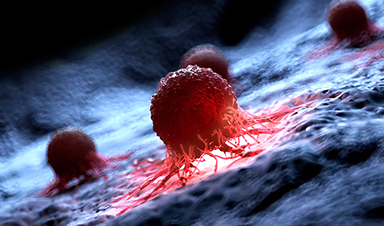
Cancer Mystery Solved: Scientists Discover How Melanoma Becomes “Immortal”
Scientists have uncovered a previously overlooked mechanism that may help melanoma cells become effectively “immortal.” Cancer cells face a major problem before they can become deadly: They have to figure out how to stop [...]
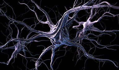
How Visual Neurons Organize Thousands of Synaptic Inputs
Summary: A new study uncovered the organizational rules that determine how neurons in the primary visual cortex process information. By imaging both the cell bodies (soma) and the individual synapses (on dendritic spines) of [...]
Scientists Just Found a Surprising Way To Destroy “Forever Chemicals”
Scientists have uncovered a new mechanism that may help break down highly persistent PFAS pollutants. PFAS have earned the nickname “forever chemicals” for a reason. These industrial compounds are so chemically durable that they [...]

Scientists Discover Cheap Material That Kills Deadly Superbugs
A new sulfur-rich antimicrobial polymer shows strong effectiveness against fungal and bacterial pathogens and may offer an affordable solution to antimicrobial resistance. Antimicrobial resistance is creating growing challenges for both healthcare and food production, [...]

What to Know About Cicada, or BA.3.2, the Latest SARS-CoV-2 Variant Under Monitoring
Like periodical cicadas, the insects for which it is nicknamed, SARS-CoV-2 Omicron subvariant BA.3.2 is only just beginning to emerge after lying low for an extended period since it first appeared. Although it was [...]

Scientists Say This Simple Supplement May Actually Reverse Heart Disease
Scientists in Japan say a common supplement may actually help “unclog” certain diseased heart arteries from the inside out. A simple food supplement sold in Japan may have helped reverse a dangerous form of [...]

New breakthrough against radiation: Korean Scientists create revolutionary shield with nanotechnology
Korean Scientists develop new nanotechnology material capable of reducing radiation impacts in space missions, hospitals, and power plants. The search for more efficient protection technologies in extreme environments has just gained an important advance. Korean [...]

Scientists Just Discovered the Hidden Trick That Keeps Your Cells Alive
A strange bead-like motion inside cells may be the secret to keeping their DNA—and health—in balance. Mitochondria are often described as the power plants of the cell because they produce the energy cells need [...]
Scientists Discover Stem Cells That Could Regrow Teeth and Bone
Scientists just uncovered the cellular “blueprint” that could one day let us regrow real teeth. Researchers at Science Tokyo have uncovered two distinct stem cell lineages that play a central role in forming tooth [...]

Scientists Uncover Fatal Weakness in “Zombie Cells” Linked to Cancer
A newly identified weakness in “zombie” cells may open the door to more precise cancer treatments by turning their own survival strategy against them. A new class of drugs takes advantage of a recently [...]

Bowel and Ovarian Cancers Are Dramatically Rising in Young Adults, Scientists Aren’t Sure Why
Cancer incidence is increasing, especially among younger adults, and current risk factors don’t fully account for the trend. Scientists suggest other underlying causes may be contributing. Cancer patterns in England are shifting in a [...]

New Immune Pathway Could Supercharge mRNA Cancer Vaccines
A surprising backup system in the immune response to mRNA vaccines may hold the key to more effective cancer treatments. The arrival of mRNA vaccines against SARS-CoV-2 in 2020 marked a turning point in the COVID-19 pandemic. Today, [...]
Scientists Discover “Molecular Switch” That Fuels Alzheimer’s Brain Inflammation
A newly identified trigger of brain inflammation could offer a fresh target for slowing Alzheimer’s progression. The brain has its own built-in immune system that identifies threats and responds to them. In Alzheimer’s disease, growing evidence [...]

Molecular Manufacturing: The Future of Nanomedicine – New book from NanoappsMedical Inc.
This book explores the revolutionary potential of atomically precise manufacturing technologies to transform global healthcare, as well as practically every other sector across society. This forward-thinking volume examines how envisaged Factory@Home systems might enable the cost-effective [...]

Forgotten Medicinal Plant Shows Promise in Fighting Dangerous Superbugs
A traditional medicinal plant, tormentil, shows promise against antibiotic-resistant bacteria in laboratory tests. Its compounds work by limiting bacterial growth and boosting antibiotic performance. Before the development of modern antibiotics, plant-based remedies were commonly [...]