AI algorithms outperformed traditional clinical risk models in a large-scale study, predicting five-year breast cancer risk more accurately. These models use mammograms as the single data source, offering potential advantages in individualizing patient care and enhancing prediction efficiency.
In a large study of thousands of mammograms, artificial intelligence (AI) algorithms outperformed the standard clinical risk model for predicting the five-year risk for breast cancer. The results of the study were published in Radiology, a journal of the Radiological Society of North America (RSNA).
A woman’s risk of breast cancer is typically calculated using clinical models such as the Breast Cancer Surveillance Consortium (BCSC) risk model, which uses self-reported and other information on the patient—including age, family history of the disease, whether she has given birth, and whether she has dense breasts—to calculate a risk score.
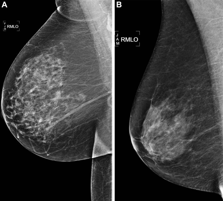
Right medial lateral oblique (RMLO) screening mammograms show negative results from 2016 in (A) a 73-year-old woman with Mirai artificial intelligence (AI) risk score with more than 90th percentile risk who developed right breast cancer in 2021 at 5 years of follow-up and (B) a 73-year-old woman with Mirai AI risk score with less than 10th percentile risk who did not develop cancer at 5 years after 5 years of follow-up. Credit: Radiological Society of North America
“We selected from the entire year of screening mammograms performed in 2016, so our study population is representative of communities in Northern California,” Dr. Arasu said.
The researchers divided the five-year study period into three time periods: interval cancer risk, or incident cancers diagnosed between 0 and 1 years; future cancer risk, or incident cancers diagnosed from between one and five years; and all cancer risk, or incident cancers diagnosed between 0 and 5 years.
Using the 2016 screening mammograms, risk scores for breast cancer over the five-year period were generated by five AI algorithms, including two academic algorithms used by researchers and three commercially available algorithms. The risk scores were then compared to each other and to the BCSC clinical risk score.
“All five AI algorithms performed better than the BCSC risk model for predicting breast cancer risk at 0 to 5 years,” Dr. Arasu said. “This strong predictive performance over the five-year period suggests AI is identifying both missed cancers and breast tissue features that help predict future cancer development. Something in mammograms allows us to track breast cancer risk. This is the ‘black box’ of AI.”
“[AI] is a tool that could help us provide personalized, precision medicine on a national level..” — Vignesh A. Arasu, M.D., Ph.D.
Some of the AI algorithms excelled at predicting patients at high risk of interval cancer, which is often aggressive and may require a second reading of mammograms, supplementary screening, or short-interval follow-up imaging. When evaluating women with the highest 10% risk as an example, AI predicted up to 28% of cancers compared to 21% predicted by BCSC.
Even AI algorithms trained for short time horizons (as low as 3 months) were able to predict the future risk of cancer up to five years when no cancer was clinically detected by screening mammography. When used in combination, the AI and BCSC risk models further improved cancer prediction.
“We’re looking for an accurate, efficient and scalable means of understanding a women’s breast cancer risk,” Dr. Arasu said. “Mammography-based AI risk models provide practical advantages over traditional clinical risk models because they use a single data source: the mammogram itself.”
Dr. Arasu said some institutions are already using AI to help radiologists detect cancer on mammograms. A person’s future risk score, which takes seconds for AI to generate, could be integrated into the radiology report shared with the patient and their physician.
“AI for cancer risk prediction offers us the opportunity to individualize every woman’s care, which isn’t systematically available,” he said. “It’s a tool that could help us provide personalized, precision medicine on a national level.”
Reference: “Comparison of Mammography AI Algorithms with a Clinical Risk Model for 5-year Breast Cancer Risk Prediction: An Observational Study” by Vignesh A. Arasu, Laurel A. Habel, Ninah S. Achacoso, Diana S. M. Buist, Jason B. Cord, Laura J. Esserman, Nola M. Hylton, M. Maria Glymour, John Kornak, Lawrence H. Kushi, Donald A. Lewis, Vincent X. Liu, Caitlin M. Lydon, Diana L. Miglioretti, Daniel A. Navarro, Albert Pu, Li Shen, Weiva Sieh, Hyo-Chun Yoon and Catherine Lee, 6 June 2023, Radiology.
DOI: 10.1148/radiol.222733
News

Younger Generations Are Aging Faster – and It May Be Fueling a Surge in Cancer
Younger generations may be aging biologically faster than those before them, and that shift could help explain rising rates of cancer at younger ages. For decades, cancer was viewed largely as a disease of [...]

Using Cannabis Could Raise Your Stroke Risk by 37%, Massive Study Reveals
Large-scale evidence suggests cannabis, cocaine, and amphetamines may directly raise stroke risk, including in younger adults. As recreational drug use becomes increasingly common, researchers are uncovering evidence that its health consequences may extend far beyond [...]

Could Vitamin C Be the Secret to Keeping Your Brain Younger?
Lower vitamin C levels were linked to reduced brain volume and weaker neural connectivity in older adults, suggesting a potential connection between nutrition and brain health. Could a common vitamin help preserve the brain [...]

This Deadly Disease Was Wiping Out Humans 5,500 Years Ago
A new study suggests plague was already a deadly threat 5,500 years ago, striking small hunter-gatherer communities long before cities and agriculture emerged. For centuries, plague has been remembered as the disease that devastated [...]

China closing in but US leads in biotech quality, commercial reach, survey finds
SAN DIEGO, June 22 (Reuters) - China, which now conducts more clinical drug trials, opens new tab than the U.S., still lags in the quality and commercial reach of its biomedical science, according to a recent survey, opens new [...]

New method generates renewable supply of progenitor immune cells
In a paper published in Cell, a USC Stem Cell-led team reports a new way of generating a renewable and expandable supply of the progenitor cells that give rise to macrophages. These immune cells help [...]

Scientists Just Discovered a Cellular Survival System That Was Never Supposed To Exist
A surprising backup pathway allows cells to make a crucial amino acid when their primary machinery fails. For decades, biologists believed cells had only one way to access a molecule they cannot live without. New [...]

Artificial cells gain porous membranes, enabling lab reactions and drug release
Artificial cells created in the laboratory offer a wide range of potential applications. Until now, however, their membranes—unlike those of real cells—have been virtually impermeable. Researchers at the Max Planck Institute for Polymer Research, [...]

Popular Weight-Loss Drugs Like Ozempic Linked to Lower Breast Cancer Risk
Ozempic and similar weight-loss drugs were linked to a striking 30% reduction in breast cancer risk in a study of more than 110,000 women. Popular weight-loss and diabetes medications such as Ozempic, Wegovy, Mounjaro, [...]

Stanford Scientists Discover Explosive New Type of Immune Cell
Scientists studying the remarkable regenerative abilities of planarian flatworms have uncovered a previously unknown type of immune cell with an unusually destructive defense strategy. What if an immune cell could wipe out nearby threats [...]

Big Pharma-backed SonoThera sounds off with $125M series B for bubble-based genetic delivery
Bay Area biotech SonoThera is bubbling to a clinical boil after raising a $125 million series B with the backing of some of the biggest names in pharma. Vida Ventures led the raise, with the venture [...]

Joint initiative of 5 EU countries calls for ‘unified approach’ to pharma framework amid US drug pricing pressure
With drug pricing pressure building from the U.S., a healthcare-focused consortium of five European countries is calling for a “unified approach” to strengthen Europe’s pharmaceutical framework and access to innovative medicines. Belgium, the Netherlands, [...]

Our books now available worldwide!
Online Sellers other than Amazon, Routledge, and IOPP Indigo Global Health Care Equivalency in the Age of Nanotechnology, Nanomedicine and Artifcial Intelligence Global Health Care Equivalency In The Age Of Nanotechnology, Nanomedicine And Artificial [...]

Molecular Manufacturing: The Future of Nanomedicine – New book from NanoappsMedical Inc.
This book explores the revolutionary potential of atomically precise manufacturing technologies to transform global healthcare, as well as practically every other sector across society. This forward-thinking volume examines how envisaged Factory@Home systems might enable the cost-effective [...]

NanoMedical Brain/Cloud Interface – Explorations and Implications. A new book from Frank Boehm
New book from Frank Boehm, NanoappsMedical Inc Founder: This book explores the future hypothetical possibility that the cerebral cortex of the human brain might be seamlessly, safely, and securely connected with the Cloud via [...]

New book from Nanoappsmedical Inc. – Global Health Care Equivalency
A new book by Frank Boehm, NanoappsMedical Inc. Founder. This groundbreaking volume explores the vision of a Global Health Care Equivalency (GHCE) system powered by artificial intelligence and quantum computing technologies, operating on secure [...]