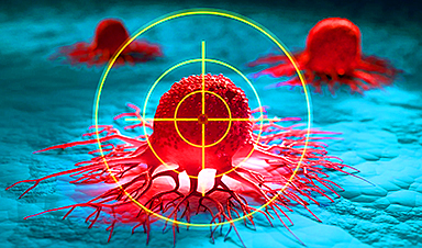
A recent study published in Small addresses the persistent difficulty of treating refractory melanoma, an aggressive form of skin cancer that often does not respond to existing therapies.
Although diagnostic tools and immunotherapies have improved in recent years, a substantial number of patients remain unresponsive to current treatment options, highlighting the need for alternative therapeutic approaches.
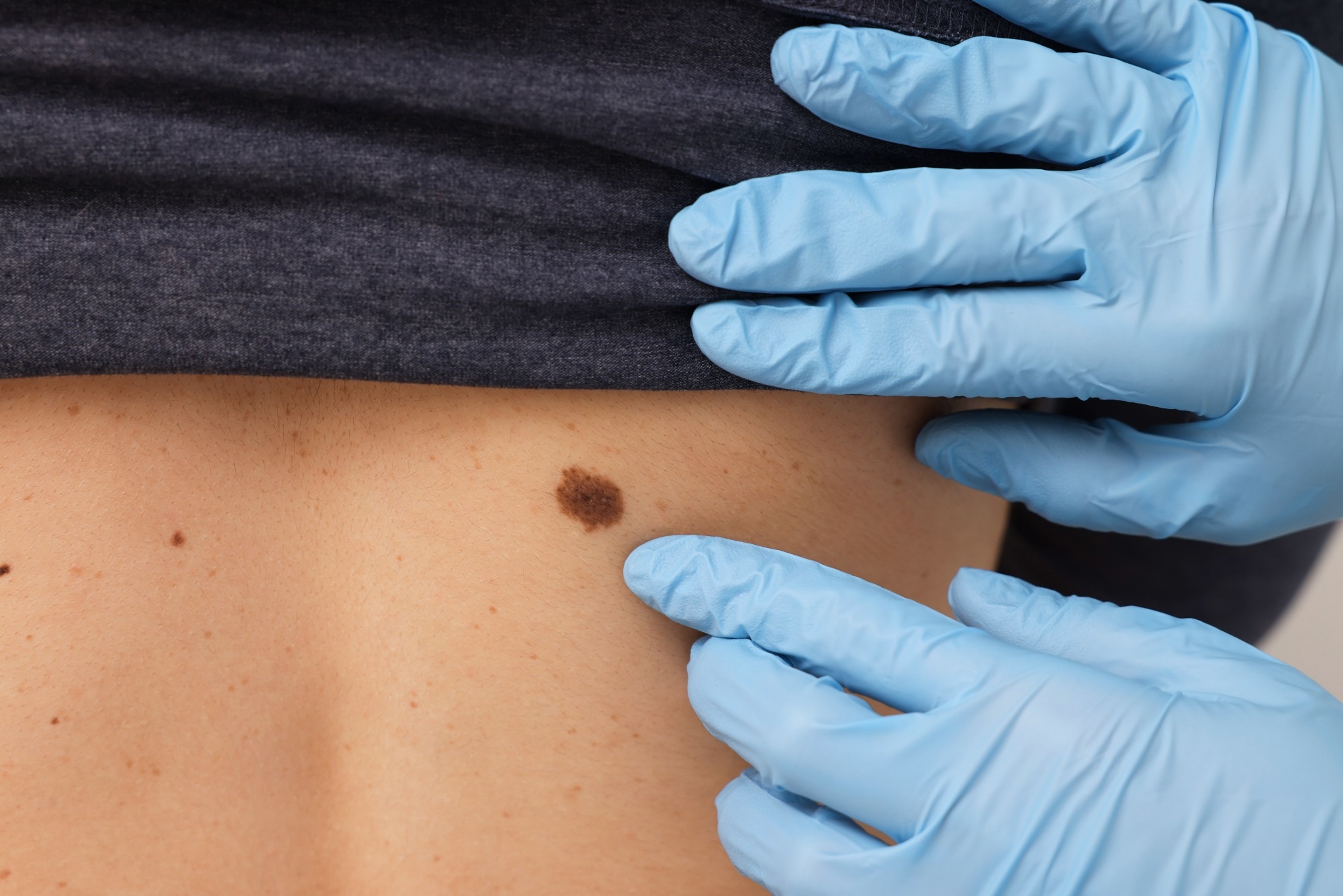
Image Credit: New Africa/Shutterstock.com
The researchers in this study explore a strategy that combines intracellular stress targeting with immune modulation.
Specifically, they investigate the co-administration of two hydrophobic drugs: copper diethyldithiocarbamate (CuET), which inhibits the p97-UFD1-NPL4 protein complex to induce endoplasmic reticulum (ER) stress and promote cytotoxicity; and 6-bromo-indirubin-3′-oxime (BIO), a GSK3 inhibitor that can influence inflammatory pathways and tumor cell growth.
Background
Melanoma becomes particularly difficult to treat once it develops resistance to standard therapies. Tumor cells can avoid immune detection and resist cell death mechanisms, reducing the effectiveness of many treatments. This study focuses on targeting both cellular stress pathways and immune checkpoints as a dual approach.
CuET disrupts protein degradation by inhibiting the p97-UFD1-NPL4 complex, leading to ER stress and apoptosis, especially in cancer cells already under stress. BIO, as a GSK3 inhibitor, affects β-catenin signaling and the production of inflammatory cytokines, which can help reshape the tumor microenvironment to enhance immune recognition.
Because both CuET and BIO are hydrophobic, systemic delivery is a challenge. To address this, the researchers developed liposome-polymer nanoparticles (LPNs) capable of encapsulating the drugs, improving their solubility, delivery precision, and release control.
The Current Study
The research included both in vitro and in vivo experiments to evaluate the drug delivery system. The team first established the optimal molar ratio of CuET to BIO using several melanoma cell lines, including B16F10 and YUMM1.7, along with their variants.
The drugs were co-loaded into LPNs made from phospholipids and stabilized with poly(vinylpyrrolidone), which improved their compatibility in aqueous environments. Particle size, surface charge, encapsulation efficiency, and stability were analyzed using dynamic light scattering and electron microscopy.
Cellular uptake and cytotoxicity were assessed using viability assays (including the sulforhodamine B method) in both two-dimensional cell cultures and three-dimensional tumor spheroids. Additional analyses (such as immunofluorescence, Western blotting, and flow cytometry) were used to track changes in β-catenin levels, immune marker expression, and T cell activation.
In vivo, the LPNs were tested in mouse models of melanoma, again using the B16F10 and YUMM1.7 cell lines, which exhibit features of therapy-resistant disease. Tumor growth, metastasis, and treatment-related toxicity were monitored through imaging, histological evaluation, and blood analysis.
Results and Discussion
The co-loaded nanoparticles demonstrated consistent particle size (100–150 nm), high encapsulation efficiency, and stability under physiological conditions. In vitro, the combination therapy showed a greater reduction in melanoma cell viability than either drug alone, indicating a synergistic cytotoxic effect.
One notable finding was BIO’s ability to counteract the accumulation of β-catenin induced by CuET. This suggests that the drug pair can modulate intracellular signaling in a way that may limit tumor proliferation and reduce metastatic potential. The combination also increased markers of ER stress and apoptosis, supporting the idea that the two drugs operate through complementary mechanisms.
Beyond direct effects on tumor cells, the study also examined the immune-related impact of the treatment. The combination therapy led to reduced expression of PD-L1 on tumor cells, potentially improving immune cell recognition. Flow cytometry revealed increased levels of immune activation markers such as CD69, along with changes in PD-1 expression on T cells. CuET alone increased PD-1 levels, a response that was moderated by the addition of BIO.
CuET was also found to suppress IL-2 secretion from activated T cells, directly influencing immune cell function. These results suggest that the therapy engages both tumor-intrinsic and immune-modulatory pathways, contributing to a more comprehensive anti-tumor response.
In vivo, treatment with the liposome-polymer nanoparticles led to a significant decrease in tumor size—about 47 % in B16F10 models and over 75 % in YUMM1.7 models. Importantly, this effect was achieved without significant toxicity. Mice maintained stable body weight, and blood and histological analyses showed no signs of liver or kidney damage.
Overall, the findings support the use of this nanocarrier system for delivering hydrophobic drug combinations, offering effective tumor suppression with a favorable safety profile.
Conclusion
This study presents a liposome-polymer nanoparticle system designed to deliver CuET and BIO in combination as a potential treatment for resistant melanoma. The formulation demonstrated stability, effective tumor suppression in vitro and in vivo, and a favorable safety profile.
By targeting ER stress, β-catenin signaling, and immune checkpoint pathways, the approach offers a multi-faceted therapeutic option for melanoma that has not responded to existing treatments.
Further research may explore the use of similar delivery systems for other drug combinations, particularly in cancers where treatment resistance remains a significant challenge.
Journal Reference
Paun R. A., et al. (2025). Liposome-Polymer Nanoparticles Loaded with Copper Diethyldithiocarbamate and 6-Bromo-Indirubin-3′-Oxime Enable the Treatment of Refractive Melanoma. Small, DOI: 10.1002/smll.202409012, https://onlinelibrary.wiley.com/doi/10.1002/smll.202409012
News

New book from NanoappsMedical Inc – Molecular Manufacturing: The Future of Nanomedicine
This book explores the revolutionary potential of atomically precise manufacturing technologies to transform global healthcare, as well as practically every other sector across society. This forward-thinking volume examines how envisaged Factory@Home systems might enable the cost-effective [...]
Scientists Discover Simple Saliva Test That Reveals Hidden Diabetes Risk
Researchers have identified a potential new way to assess metabolic health using saliva instead of blood. High insulin levels in the blood, known as hyperinsulinemia, can reveal metabolic problems long before obvious symptoms appear. It is [...]

One Nasal Spray Could Protect Against COVID, Flu, Pneumonia, and More
A single nasal spray vaccine may one day protect against viruses, pneumonia, and even allergies. For decades, scientists have dreamed of creating a universal vaccine capable of protecting against many different pathogens. The idea [...]
New AI Model Predicts Cancer Spread With Incredible Accuracy
Scientists have developed an AI system that analyzes complex gene-expression signatures to estimate the likelihood that a tumor will spread. Why do some tumors spread throughout the body while others remain confined to their [...]

Scientists Discover DNA “Flips” That Supercharge Evolution
In Lake Malawi, hundreds of species of cichlid fish have evolved with astonishing speed, offering scientists a rare opportunity to study how biodiversity arises. Researchers have identified segments of “flipped” DNA that may allow fish to adapt rapidly [...]

Our books now available worldwide!
Online Sellers other than Amazon, Routledge, and IOPP Indigo Global Health Care Equivalency in the Age of Nanotechnology, Nanomedicine and Artifcial Intelligence Global Health Care Equivalency In The Age Of Nanotechnology, Nanomedicine And Artificial [...]
Scientists Discover Why Some COVID Survivors Still Can’t Taste Food Years Later
A new study provides the first direct biological evidence explaining why some people continue to experience taste loss long after recovering from COVID-19. Researchers have uncovered specific biological changes in taste buds that could help [...]
Catching COVID significantly raises the risk of developing kidney disease, researchers find
Catching Covid significantly raises the risk of developing deadly kidney disease, research has shown. The virus was found to increase the chances that patients will develop the incurable condition by around 50 per cent. [...]
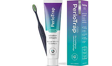
New Toothpaste Stops Gum Disease Without Harming Healthy Bacteria
Researchers have developed a targeted approach to combat periodontitis without disrupting the natural balance of the oral microbiome. The innovation could reshape how gum disease is treated while preserving beneficial bacteria. The human mouth [...]

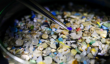
Plastic Without End: Are We Polluting the Planet for Eternity?
The Kunming Montreal Global Biodiversity Framework calls for the elimination of plastic pollution by 2030. If that goal has been clearly set, why have meaningful measures that create real change still not been implemented? [...]
Scientists Rewire Natural Killer Cells To Attack Cancer Faster and Harder
Researchers tested new CAR designs in NK-92 cells and found the modified cells killed tumor cells more effectively, showing stronger anti-cancer activity. Researchers at the Ribeirão Preto Blood Center and the Center for Cell-Based [...]
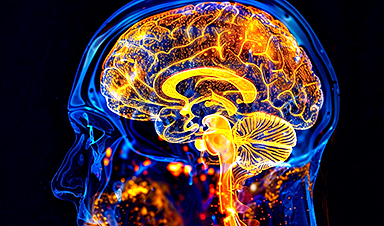
New “Cellular” Target Could Transform How We Treat Alzheimer’s Disease
A new study from researchers highlights an unexpected player in Alzheimer’s disease: aging astrocytes. Senescent astrocytes have been identified as a major contributor to Alzheimer’s progression. The cells lose protective functions and fuel inflammation, particularly in [...]

Treating a Common Dental Infection… Effects That Extend Far Beyond the Mouth
Successful root canal treatment may help lower inflammation associated with heart disease and improve blood sugar and cholesterol levels. Treating an infected tooth with a successful root canal procedure may do more than relieve [...]
Microplastics found in prostate tumors in small study
In a new study, researchers found microplastics deep inside prostate cancer tumors, raising more questions about the role the ubiquitous pollutants play in public health. The findings — which come from a small study of 10 [...]
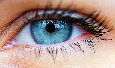
All blue-eyed people have this one thing in common
All Blue-Eyed People Have This One Thing In Common Blue Eyes Aren’t Random—Research Traces Them Back to One Prehistoric Human It sounds like a myth at first — something you’d hear in a folklore [...]
Scientists reveal how exercise protects the brain from Alzheimer’s
Researchers at UC San Francisco have identified a biological process that may explain why exercise sharpens thinking and memory. Their findings suggest that physical activity strengthens the brain's built in defense system, helping protect [...]