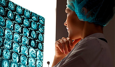
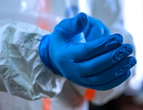
In today’s hospitals and healthcare clinics, a new doctor’s new assistant is now often on the job — in the form of artificial intelligence. Whether it’s analyzing medical images or guiding robots that assist with surgeries, AI is making steady inroads into our hospitals and clinics. Need an online nursing assistant or a watchdog that helps detect dosage errors? There’s an AI application for that.
The advent of AI in healthcare is a promising trend in terms of both patient care and economic efficiency. AI can help us address a forecasted shortage of physicians, particularly in specialty-care fields, while containing the costs of caring for an aging and growing population. A recent study by a team of researchers from the consulting firm Accenture found that the use of 10 promising AI applications could create up to $150 billion in annual savings for U.S. healthcare by 2026.
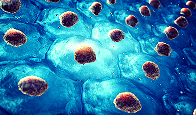
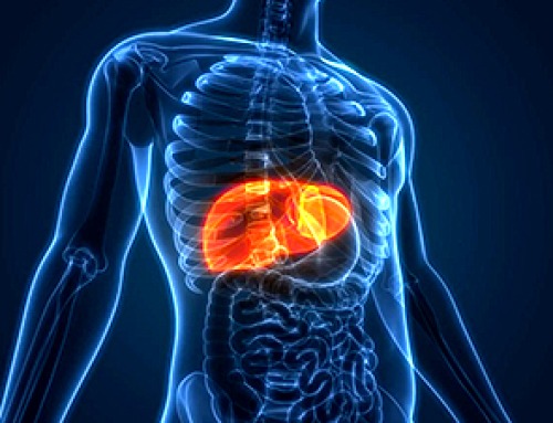
For an example of the potential of AI in healthcare, we need to look no further than Gustave Roussy, a leading European center for cancer research and care. In a study published this summer in the medical journal The Lancet Oncology, a team of medical researchers from Gustave Roussy and a few other institutions demonstrated that AI can process medical images to extract biological and clinical information to help with immunotherapy treatment, according to a news release on the study.
In this groundbreaking study, the researchers used an algorithm they designed and developed to analyze CT scan images and create a “radiomic signature.” This signature defines the level of lymphocyte infiltration of a tumor — or the degree to which immune cells have moved from the blood into a tumor cell. The radiomic signature also provides a predictive score for the efficacy of immunotherapy in the patient.
Here’s how AI played into this research: Using an approach based on machine learning, the team first taught the algorithm to use relevant information extracted from CT scans of patients participating in the study. Then, based solely on images, the algorithm learned to predict what the genome might have revealed about the tumor immune infiltrate, and it established the radiomic signature.
The announcement summarizing the findings of the study notes that in the future, physicians might be able to use imaging to identify biological phenomena in a tumor located in any part of the body without having to perform a biopsy.
At Dell EMC, this research is particularly close to our hearts because we are active supporters of Gustave Roussy.
Image Credit: Dell EMC
News This Week
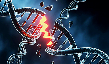
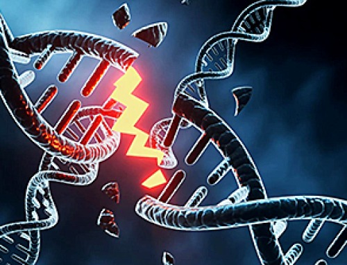
Building the Brain Requires Millions of Dangerous DNA Breaks
Scientists discovered that building a healthy brain involves an unexpected step: young neurons routinely break and rapidly repair their own DNA. As the brain develops, newly formed nerve cells must travel through tightly packed tissue [...]
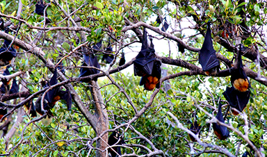
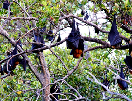
One Tiny Change May Explain How Viruses Jump From Bats to Humans
Scientists found that one tiny genetic change may determine whether a bat virus stays in bats or becomes a human threat. Most infectious disease outbreaks begin when a virus or other pathogen crosses from animals into [...]
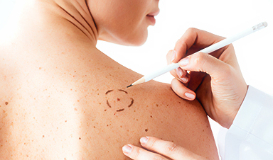
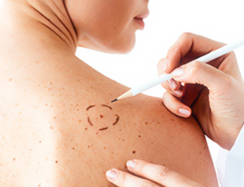
Scientists Discover 250+ Genes That Could Lead to New Ways To Prevent Melanoma
The world’s largest study of mole genetics identified hundreds of genes tied to melanoma risk, uncovering potential new drug targets and paving the way for more accurate melanoma screening and prevention. Researchers at QIMR [...]
Breakthrough Diabetes Treatment Reprograms the Immune System
An engineered stem cell therapy reversed new-onset Type 1 diabetes in mice by shifting the immune system away from attacking insulin-producing cells. For more than a century, people with Type 1 diabetes have relied [...]
Taking the world’s temperature: WHO chief spotlights global health emergencies
Taking the world’s temperature on pressing health matters, WHO Director-General Tedros Adhanom Ghebreyesus provided the latest on current global challenges - and successes when it comes to international cooperation. “The outbreaks of hantavirus, Ebola and Marburg all show [...]
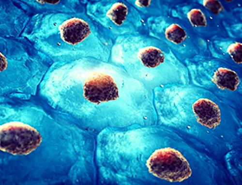
Scientists Create Tiny “Mini Livers” That Could One Day Replace Liver Transplants
Engineered tissue grafts could help perform key liver functions and benefit thousands of people living with liver failure. The liver is one of the body’s hardest-working organs, carrying out hundreds of vital jobs, from [...]

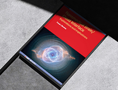
NanoMedical Brain/Cloud Interface – Explorations and Implications. A new book from Frank Boehm
New book from Frank Boehm, NanoappsMedical Inc Founder: This book explores the future hypothetical possibility that the cerebral cortex of the human brain might be seamlessly, safely, and securely connected with the Cloud via [...]
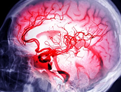
Scientists Discover Surprising Way To Help the Brain Recover After Stroke
A new study suggests that strengthening the body’s natural circadian rhythms may help the brain recover after stroke, even when treatment begins days after the injury. Every year, millions of people survive a stroke, [...]

Our books now available worldwide!
Online Sellers other than Amazon, Routledge, and IOPP Indigo Global Health Care Equivalency in the Age of Nanotechnology, Nanomedicine and Artifcial Intelligence Global Health Care Equivalency In The Age Of Nanotechnology, Nanomedicine And Artificial [...]

Younger Generations Are Aging Faster – and It May Be Fueling a Surge in Cancer
Younger generations may be aging biologically faster than those before them, and that shift could help explain rising rates of cancer at younger ages. For decades, cancer was viewed largely as a disease of [...]

Using Cannabis Could Raise Your Stroke Risk by 37%, Massive Study Reveals
Large-scale evidence suggests cannabis, cocaine, and amphetamines may directly raise stroke risk, including in younger adults. As recreational drug use becomes increasingly common, researchers are uncovering evidence that its health consequences may extend far beyond [...]

Could Vitamin C Be the Secret to Keeping Your Brain Younger?
Lower vitamin C levels were linked to reduced brain volume and weaker neural connectivity in older adults, suggesting a potential connection between nutrition and brain health. Could a common vitamin help preserve the brain [...]

This Deadly Disease Was Wiping Out Humans 5,500 Years Ago
A new study suggests plague was already a deadly threat 5,500 years ago, striking small hunter-gatherer communities long before cities and agriculture emerged. For centuries, plague has been remembered as the disease that devastated [...]

China closing in but US leads in biotech quality, commercial reach, survey finds
SAN DIEGO, June 22 (Reuters) - China, which now conducts more clinical drug trials, opens new tab than the U.S., still lags in the quality and commercial reach of its biomedical science, according to a recent survey, opens new [...]

New method generates renewable supply of progenitor immune cells
In a paper published in Cell, a USC Stem Cell-led team reports a new way of generating a renewable and expandable supply of the progenitor cells that give rise to macrophages. These immune cells help [...]

Scientists Just Discovered a Cellular Survival System That Was Never Supposed To Exist
A surprising backup pathway allows cells to make a crucial amino acid when their primary machinery fails. For decades, biologists believed cells had only one way to access a molecule they cannot live without. New [...]
Leave A Comment