| Nanoparticles are everywhere. They are in our body as protein aggregates, lipid vesicles, or viruses. They are in our drinking water in the form of impurities. They are in the air we breath as pollutants. At the same time, many drugs are based on the delivery of nanoparticles, including the vaccines we have been recently given. | |
| Keeping with the pandemics, quick tests used for the detection the SARS-Cov-2 are based on nanoparticles too. The red line, which we monitor day by day, contains myriads of gold nanoparticles coated with antibodies against proteins that report infection. | |
| Technically, one calls something a nanoparticle when its size (diameter) is smaller than one micrometer (one thousandth of a millimeter). Objects of the order of one micrometer can still be measured in a normal microscope, but particles that are much smaller, say smaller than 0.2 micrometers, become exceedingly difficult to measure or characterize. Interestingly, this is also the size range of viruses, which can become as small as 0.02 micrometers. |
| Over the years, scientists and engineers have devised a number of instruments for characterizing nanoparticles. Ideally, one wants to measure their concentration, assess their size and size distribution, and determine their substance. A high-end example is an electron microscope. | |
| But this technology has many shortcomings. It is very bulky and expensive, and the studies take too long because samples have to be carefully prepared and be put into vacuum. And even then, it remains difficult to determine the substance of the particles one sees in an electron microscope. | |
| A quick, reliable, light and portable device that can be used in the doctor’s office or in the field would have a huge impact. A few optical instruments on the market offer such solutions, but their resolution and precision have been insufficient for examining smaller nanoparticles, e.g., much smaller than 0.1 micrometer (or otherwise said 100 nm). | |
| A group of researchers at the Max Planck Institute for the Science of Light and Max-Planck-Zentrum für Physik und Medizin have now invented a new device that offers a big leap in the characterization of nanoparticles. The method is called iNTA, short for Interferometric Nanoparticle Tracking Analysis. | |
| Their results are reported in the May issue of Nature Methods (“Precision size and refractive index analysis of weakly scattering nanoparticles in polydispersions”). |
| The method is based on the interferometric detection of the light scattered by individual nanoparticles that wander around in a liquid. In such a medium, thermal energy perpetually moves particles in random directions. It turns out that the space that a particle explores in a given time correlates with its size. In other words, small particles move “faster” and cover a bigger volume than large particles. | |
| The equation that describes this phenomenon – the Stokes-Einstein relation – dates back to the beginning of the last century and since then has found use in many applications. In a nutshell, if one could follow a nanoparticle and collect statistics about its jittery trajectory, one could deduce its size. So, the challenge is to record very fast movies of tiny particles wizzing by. | |
| Scientists at MPL have developed a special microscopy method over the past two decades, known as interferometric scattering (iSCAT) microscopy. This technique is extremely sensitive in detecting nanoparticles. By applying iSCAT to the problem of diffusing nanoparticles, the MPL group realized that they can outperform the existing instruments on the market. The new technology has a particular edge in deciphering mixtures of nanoparticles with different sizes and different materials. | |
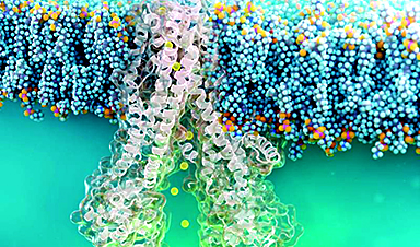
| The applications of the new method are manifold. A particularly exciting line of applications concerns nano-sized vehicles that are secreted from cells, the so-called extracellular vesicles. These are made of a lipid shell, much like a nano soap bubble. But the shell and the inner liquid also contain proteins, which tell us about the origin of the vesicles, i.e. from which organ or cellular process. When the protein amount and/or the vesicle size deviate from the normal range, it could be that the person is ill. Therefore, it is very important to find ways to characterize extracellular vesicles. | |
| The researchers at the MPL and MPZPM are now working on developing a bench-top system to enable scientists worldwide to benefit from the advantages of iNTA. |
News

New Immune Pathway Could Supercharge mRNA Cancer Vaccines
A surprising backup system in the immune response to mRNA vaccines may hold the key to more effective cancer treatments. The arrival of mRNA vaccines against SARS-CoV-2 in 2020 marked a turning point in the COVID-19 pandemic. Today, [...]
Scientists Discover “Molecular Switch” That Fuels Alzheimer’s Brain Inflammation
A newly identified trigger of brain inflammation could offer a fresh target for slowing Alzheimer’s progression. The brain has its own built-in immune system that identifies threats and responds to them. In Alzheimer’s disease, growing evidence [...]

Molecular Manufacturing: The Future of Nanomedicine – New book from NanoappsMedical Inc.
This book explores the revolutionary potential of atomically precise manufacturing technologies to transform global healthcare, as well as practically every other sector across society. This forward-thinking volume examines how envisaged Factory@Home systems might enable the cost-effective [...]
Forgotten Medicinal Plant Shows Promise in Fighting Dangerous Superbugs
A traditional medicinal plant, tormentil, shows promise against antibiotic-resistant bacteria in laboratory tests. Its compounds work by limiting bacterial growth and boosting antibiotic performance. Before the development of modern antibiotics, plant-based remedies were commonly [...]

NanoMedical Brain/Cloud Interface – Explorations and Implications. A new book from Frank Boehm
New book from Frank Boehm, NanoappsMedical Inc Founder: This book explores the future hypothetical possibility that the cerebral cortex of the human brain might be seamlessly, safely, and securely connected with the Cloud via [...]

New Research Finds Shocking Link Between Chili Peppers and Cancer
If you love spicy food, you are not alone. But scientists are taking a closer look at whether eating a lot of chili peppers could affect your cancer risk. Could your love of spicy [...]

New book from Nanoappsmedical Inc. – Global Health Care Equivalency
A new book by Frank Boehm, NanoappsMedical Inc. Founder. This groundbreaking volume explores the vision of a Global Health Care Equivalency (GHCE) system powered by artificial intelligence and quantum computing technologies, operating on secure [...]
Scientists Create “Neurobots” – Living Machines With Their Own Nervous Systems
Neurobots—xenobots with neurons—show self-organized nervous systems and enhanced behaviors, revealing new insights into how biology builds functional structures. In 2020, researchers at Tufts University developed tiny living structures known as xenobots using frog cells. These microscopic organisms [...]

Our books now available worldwide!
Online Sellers other than Amazon, Routledge, and IOPP Indigo Global Health Care Equivalency in the Age of Nanotechnology, Nanomedicine and Artifcial Intelligence Global Health Care Equivalency In The Age Of Nanotechnology, Nanomedicine And Artificial [...]

Amazonian Chocolate Could Become the Next Superfood, Scientists Say
New research into Amazonian cocoa reveals that its value may extend beyond flavor alone. Chocolate from the Amazon is already known worldwide for its distinctive taste, but new research suggests it may offer even [...]
Nanobody repairs misfolded CFTR inside cells, boosting function in cystic fibrosis
A tiny antibody component could fundamentally transform the treatment of cystic fibrosis: For the first time, researchers have succeeded in developing a so-called nanobody that penetrates directly into human cells and can repair the [...]

20-Year Study Finds Daily Multivitamins Don’t Extend Lifespan
A large, decades-long study of over 390,000 U.S. adults challenges a widespread assumption about daily multivitamins. Multivitamins are a daily habit for millions of Americans, often taken with the expectation that they will extend [...]
Novel Investment Paradigms for Regenerative Healthcare Ecosystems
Introduction The transition toward regenerative healthcare ecosystems—anchored in wellness optimization, disease prevention, eradication strategies, and healthy longevity—necessitates a structural reconfiguration of capital architectures, governance models, and incentive design. Regenerative healthcare, by definition, transcends episodic [...]
What If Consciousness Exists Beyond Your Brain
Scientists still don’t know how consciousness emerges from the brain. New ideas suggest it may not emerge at all, but instead be a basic feature of reality. Is consciousness produced by the brain, or [...]
Scientists Discover Way To Treat Lung Cancer and Its Deadly Side Effect Together
A new approach using lipid nanoparticles to deliver genetic material is showing promise in tackling two major challenges in lung cancer at once.Researchers at Oregon State University have designed a new way to tackle two of [...]

Saunas Activate Your Immune System
A brief sauna session may quietly mobilize the immune system. A sauna session may do more than raise your heart rate and body temperature. A new study from Finland found that it also briefly [...]