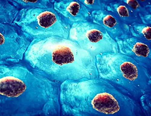
Medical diagnostics expert, doctor's assistant, and cartographer are all fair titles for an artificial intelligence model developed by researchers at the Beckman Institute for Advanced Science and Technology.
Their new model accurately identifies tumors and diseases in medical images and is programmed to explain each diagnosis with a visual map. The tool's unique transparency allows doctors to easily follow its line of reasoning, double-check for accuracy, and explain the results to patients.
"The idea is to help catch cancer and disease in its earliest stages — like an X on a map — and understand how the decision was made. Our model will help streamline that process and make it easier on doctors and patients alike," said Sourya Sengupta, the study's lead author and a graduate research assistant at the Beckman Institute.
This research appeared in IEEE Transactions on Medical Imaging.
Cats and dogs and onions and ogres
First conceptualized in the 1950s, artificial intelligence — the concept that computers can learn to adapt, analyze, and problem-solve like humans do — has reached household recognition, due in part to ChatGPT and its extended family of easy-to-use tools.
Machine learning, or ML, is one of many methods researchers use to create artificially intelligent systems. ML is to AI what driver's education is to a 15-year-old: a controlled, supervised environment to practice decision-making, calibrating to new environments, and rerouting after a mistake or wrong turn.
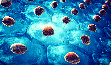
Deep learning — machine learning's wiser and worldlier relative — can digest larger quantities of information to make more nuanced decisions. Deep learning models derive their decisive power from the closest computer simulations we have to the human brain: deep neural networks.
These networks — just like humans, onions, and ogres — have layers, which makes them tricky to navigate. The more thickly layered, or nonlinear, a network's intellectual thicket, the better it performs complex, human-like tasks.
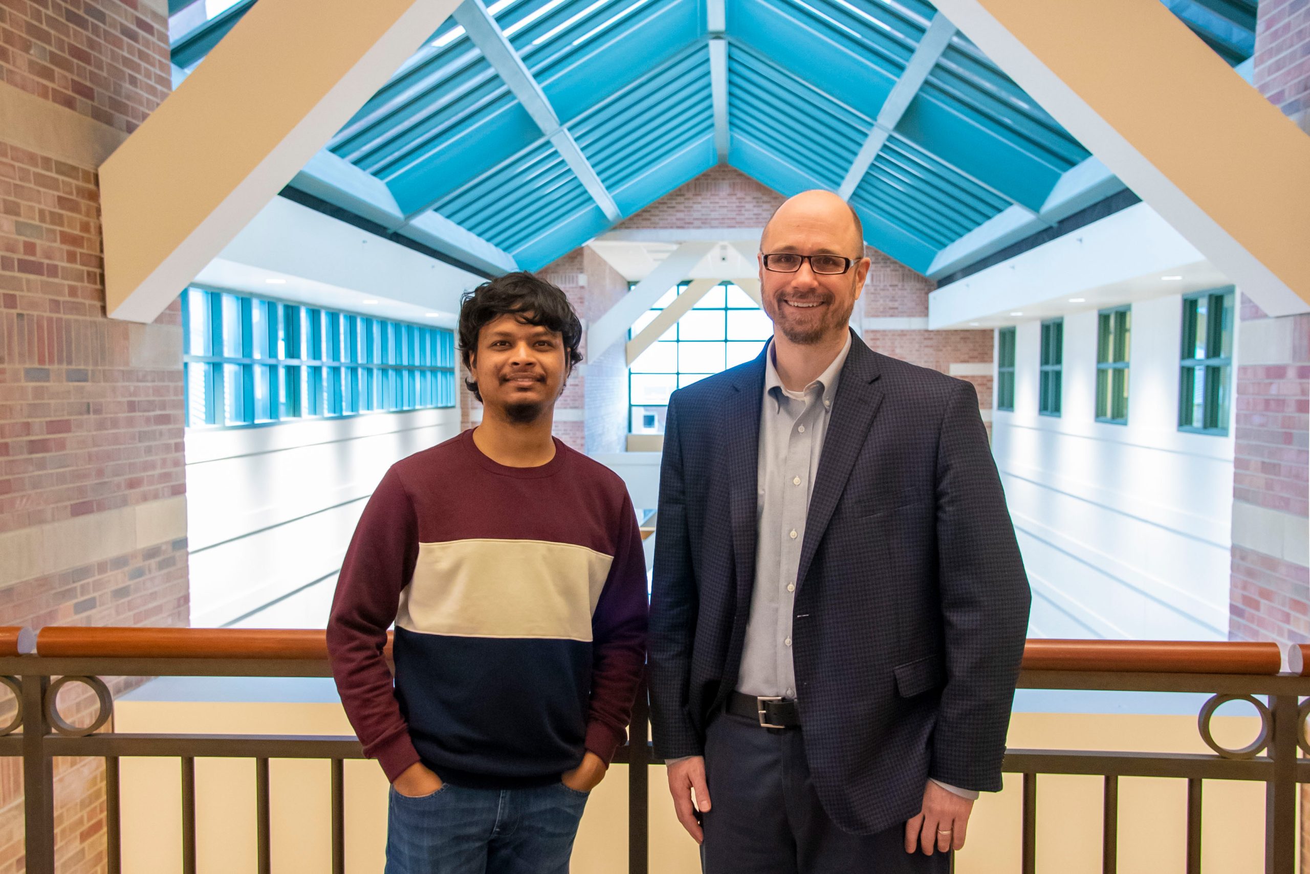
Researchers at the Beckman Institute led by Mark Anastasio (right) and Sourya Sengupta developed an artificial intelligence model that can accurately identify tumors and diseases in medical images. The tool draws a map to explain each diagnosis, helping doctors follow its line of reasoning, check for accuracy, and explain the results to patients. Credit: Jenna Kurtzweil, Beckman Institute Communications Office
Consider a neural network trained to differentiate between pictures of cats and pictures of dogs. The model learns by reviewing images in each category and filing away their distinguishing features (like size, color, and anatomy) for future reference. Eventually, the model learns to watch out for whiskers and cry Doberman at the first sign of a floppy tongue.
But deep neural networks are not infallible — much like overzealous toddlers, said Sengupta, who studies biomedical imaging in the University of Illinois Urbana-Champaign Department of Electrical and Computer Engineering.
"They get it right sometimes, maybe even most of the time, but it might not always be for the right reasons," he said. "I'm sure everyone knows a child who saw a brown, four-legged dog once and then thought that every brown, four-legged animal was a dog."
Sengupta's gripe? If you ask a toddler how they decided, they will probably tell you.
"But you can't ask a deep neural network how it arrived at an answer," he said.
The black box problem
Sleek, skilled, and speedy as they may be, deep neural networks struggle to master the seminal skill drilled into high school calculus students: showing their work. This is referred to as the black box problem of artificial intelligence, and it has baffled scientists for years.
On the surface, coaxing a confession from the reluctant network that mistook a Pomeranian for a cat does not seem unbelievably crucial. But the gravity of the black box sharpens as the images in question become more life-altering. For example: X-ray images from a mammogram that may indicate early signs of breast cancer.
The process of decoding medical images looks different in different regions of the world.
"In many developing countries, there is a scarcity of doctors and a long line of patients. AI can be helpful in these scenarios," Sengupta said.
When time and talent are in high demand, automated medical image screening can be deployed as an assistive tool — in no way replacing the skill and expertise of doctors, Sengupta said. Instead, an AI model can pre-scan medical images and flag those containing something unusual — like a tumor or early sign of disease, called a biomarker — for a doctor's review. This method saves time and can even improve the performance of the person tasked with reading the scan.
These models work well, but their bedside manner leaves much to be desired when, for example, a patient asks why an AI system flagged an image as containing (or not containing) a tumor.
Historically, researchers have answered questions like this with a slew of tools designed to decipher the black box from the outside in. Unfortunately, the researchers using them are often faced with a similar plight as the unfortunate eavesdropper, leaning against a locked door with an empty glass to their ear.
"It would be so much easier to simply open the door, walk inside the room, and listen to the conversation firsthand," Sengupta said.
To further complicate the matter, many variations of these interpretation tools exist. This means that any given black box may be interpreted in "plausible but different" ways, Sengupta said.
"And now the question is: which interpretation do you believe?" he said. "There is a chance that your choice will be influenced by your subjective bias, and therein lies the main problem with traditional methods."
Sengupta's solution? An entirely new type of AI model that interprets itself every time — that explains each decision instead of blandly reporting the binary of "tumor versus non-tumor," Sengupta said.
No water glass needed, in other words, because the door has disappeared.
Mapping the model
A yogi learning a new posture must practice it repeatedly. An AI model trained to tell cats from dogs studying countless images of both quadrupeds.
An AI model functioning as a doctor's assistant is raised on a diet of thousands of medical images, some with abnormalities and some without. When faced with something never-before-seen, it runs a quick analysis and spits out a number between 0 and 1. If the number is less than .5, the image is not assumed to contain a tumor; a numeral greater than .5 warrants a closer look.
Sengupta's new AI model mimics this setup with a twist: the model produces a value plus a visual map explaining its decision.
The map — referred to by the researchers as an equivalency map, or E-map for short — is essentially a transformed version of the original X-ray, mammogram, or other medical image medium. Like a paint-by-numbers canvas, each region of the E-map is assigned a number. The greater the value, the more medically interesting the region is for predicting the presence of an anomaly. The model sums up the values to arrive at its final figure, which then informs the diagnosis.
"For example, if the total sum is 1, and you have three values represented on the map — .5, .3, and .2 — a doctor can see exactly which areas on the map contributed more to that conclusion and investigate those more fully," Sengupta said.
This way, doctors can double-check how well the deep neural network is working — like a teacher checking the work on a student's math problem — and respond to patients' questions about the process.
"The result is a more transparent, trustable system between doctor and patient," Sengupta said.
X marks the spot
The researchers trained their model on three different disease diagnosis tasks including more than 20,000 total images.
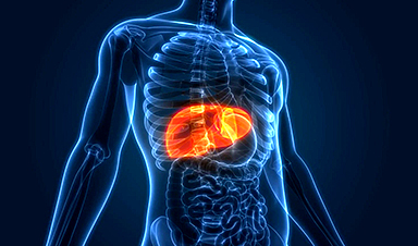
First, the model reviewed simulated mammograms and learned to flag early signs of tumors. Second, it analyzed optical coherence tomography images of the retina, where it practiced identifying a buildup called Drusen that may be an early sign of macular degeneration. Third, the model studied chest X-rays and learned to detect cardiomegaly, a heart enlargement condition that can lead to disease.
Once the mapmaking model had been trained, the researchers compared its performance to existing black-box AI systems — the ones without a self-interpretation setting. The new model performed comparably to its counterparts in all three categories, with accuracy rates of 77.8% for mammograms, 99.1% for retinal OCT images, and 83% for chest X-rays compared to the existing 77.8%, 99.1%, and 83.33.%
These high accuracy rates are a product of the deep neural network, the non-linear layers of which mimic the nuance of human neurons.
To create such a complicated system, the researchers peeled the proverbial onion and drew inspiration from linear neural networks, which are simpler and easier to interpret.
"The question was: How can we leverage the concepts behind linear models to make non-linear deep neural networks also interpretable like this?" said principal investigator Mark Anastasio, a Beckman Institute researcher and the Donald Biggar Willet Professor and Head of the Illinois Department of Bioengineering. "This work is a classic example of how fundamental ideas can lead to some novel solutions for state-of-the-art AI models."
The researchers hope that future models will be able to detect and diagnose anomalies all over the body and even differentiate between them.
"I am excited about our tool's direct benefit to society, not only in terms of improving disease diagnoses but also improving trust and transparency between doctors and patients," Anastasio said.
Reference: "A Test Statistic Estimation-based Approach for Establishing Self-interpretable CNN-based Binary Classifiers" by Sourya Sengupta and Mark A. Anastasio, 1 January 2024, IEEE Transactions on Medical Imaging.
DOI: 10.1109/TMI.2023.3348699
News

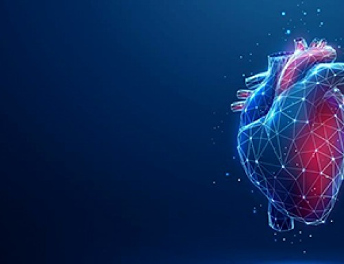
Stem cell organoids repair heart microvessels in coronary artery disease models
A Stanford University team has shown that vascular organoids derived from human stem cells can repair the heart’s microvessel network in pigs with ischaemic heart disease – a proof-of-concept advancement that could open new therapeutic [...]

Goodbye GP waiting rooms, hello prevention at home
Prevention is suddenly everywhere in NHS reform. The recent £340m community pharmacy deal is moving more services onto the high street. Community Diagnostic Centres are being expanded, and the Neighbourhood Health Framework sets out [...]
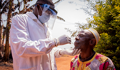
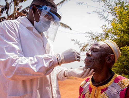
Ebola control is weakened by mistrust and cultural insensitivity
Effective response depends on cooperation with communities and frontline workers, writes Zaeem ul Haq The current Bundibugyo Ebola outbreak in the Democratic Republic of the Congo (DRC) and Uganda is exposing dangerous gaps in [...]
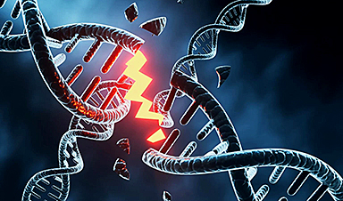
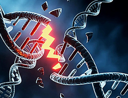
Building the Brain Requires Millions of Dangerous DNA Breaks
Scientists discovered that building a healthy brain involves an unexpected step: young neurons routinely break and rapidly repair their own DNA. As the brain develops, newly formed nerve cells must travel through tightly packed tissue [...]
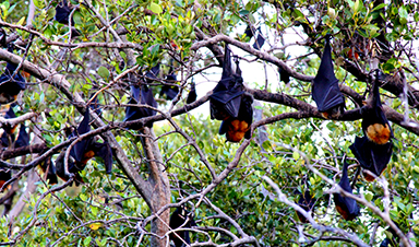
One Tiny Change May Explain How Viruses Jump From Bats to Humans
Scientists found that one tiny genetic change may determine whether a bat virus stays in bats or becomes a human threat. Most infectious disease outbreaks begin when a virus or other pathogen crosses from animals into [...]
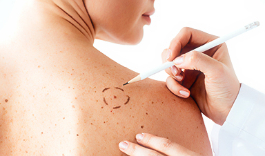
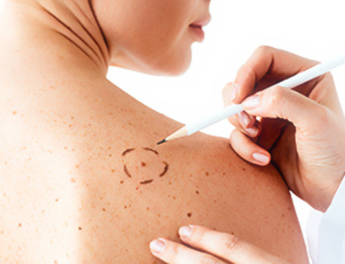
Scientists Discover 250+ Genes That Could Lead to New Ways To Prevent Melanoma
The world’s largest study of mole genetics identified hundreds of genes tied to melanoma risk, uncovering potential new drug targets and paving the way for more accurate melanoma screening and prevention. Researchers at QIMR [...]
Breakthrough Diabetes Treatment Reprograms the Immune System
An engineered stem cell therapy reversed new-onset Type 1 diabetes in mice by shifting the immune system away from attacking insulin-producing cells. For more than a century, people with Type 1 diabetes have relied [...]
Taking the world’s temperature: WHO chief spotlights global health emergencies
Taking the world’s temperature on pressing health matters, WHO Director-General Tedros Adhanom Ghebreyesus provided the latest on current global challenges - and successes when it comes to international cooperation. “The outbreaks of hantavirus, Ebola and Marburg all show [...]
Scientists Create Tiny “Mini Livers” That Could One Day Replace Liver Transplants
Engineered tissue grafts could help perform key liver functions and benefit thousands of people living with liver failure. The liver is one of the body’s hardest-working organs, carrying out hundreds of vital jobs, from [...]

NanoMedical Brain/Cloud Interface – Explorations and Implications. A new book from Frank Boehm
New book from Frank Boehm, NanoappsMedical Inc Founder: This book explores the future hypothetical possibility that the cerebral cortex of the human brain might be seamlessly, safely, and securely connected with the Cloud via [...]
Scientists Discover Surprising Way To Help the Brain Recover After Stroke
A new study suggests that strengthening the body’s natural circadian rhythms may help the brain recover after stroke, even when treatment begins days after the injury. Every year, millions of people survive a stroke, [...]

Our books now available worldwide!
Online Sellers other than Amazon, Routledge, and IOPP Indigo Global Health Care Equivalency in the Age of Nanotechnology, Nanomedicine and Artifcial Intelligence Global Health Care Equivalency In The Age Of Nanotechnology, Nanomedicine And Artificial [...]

Younger Generations Are Aging Faster – and It May Be Fueling a Surge in Cancer
Younger generations may be aging biologically faster than those before them, and that shift could help explain rising rates of cancer at younger ages. For decades, cancer was viewed largely as a disease of [...]

Using Cannabis Could Raise Your Stroke Risk by 37%, Massive Study Reveals
Large-scale evidence suggests cannabis, cocaine, and amphetamines may directly raise stroke risk, including in younger adults. As recreational drug use becomes increasingly common, researchers are uncovering evidence that its health consequences may extend far beyond [...]

Could Vitamin C Be the Secret to Keeping Your Brain Younger?
Lower vitamin C levels were linked to reduced brain volume and weaker neural connectivity in older adults, suggesting a potential connection between nutrition and brain health. Could a common vitamin help preserve the brain [...]

This Deadly Disease Was Wiping Out Humans 5,500 Years Ago
A new study suggests plague was already a deadly threat 5,500 years ago, striking small hunter-gatherer communities long before cities and agriculture emerged. For centuries, plague has been remembered as the disease that devastated [...]