A cutting-edge X-ray method reveals the 3D orientation of nanoscale material structures, offering fresh insights into their functionality.
Researchers at the Swiss Light Source (SLS) have developed a groundbreaking technique called X-ray linear dichroic orientation tomography (XL-DOT). This method reveals the three-dimensional arrangement of a material's structural building blocks at the nanoscale. Its first application focused on a polycrystalline catalyst, enabling scientists to visualize crystal grains, grain boundaries, and defects—critical features that influence catalyst performance. Beyond catalysis, XL-DOT offers unprecedented insights into the structure of various functional materials used in information technology, energy storage, and biomedical applications.
Advancements in Non-Destructive Imaging of Material Microstructures
Zooming into the micro- or nanostructure of functional materials — whether natural or man-made — reveals countless coherent domains or grains. These grains are distinct regions where molecules and atoms are arranged in orderly, repeating patterns.
The arrangement of these grains is closely tied to the material's properties. Their size, orientation, and distribution can mean the difference between a sturdy brick and a crumbling stone. They determine how ductile a metal is, how efficiently a semiconductor transfers electrons, and how well ceramics conduct heat. This structural organization also plays a critical role in biological materials; for example, collagen fibers are made of interwoven fibrils, and their alignment affects the mechanical strength of connective tissues.
These domains are often tiny: tens of nanometers in size. And it is their arrangement in three dimensions over extended volumes that is property-determining. Yet until now, techniques to probe the organization of materials at the nanoscale have largely been confined to two dimensions or are destructive in nature.
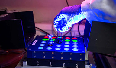
Now, using X-rays generated by the Swiss Light Source SLS, a collaborative team of researchers from Paul Scherrer Institute PSI, ETH Zurich, the University of Oxford and the Max Plank Institute for Chemical Physics of Solids have succeeded in creating an imaging technique to access this information in three-dimensions.
"We Not Only Look Inside, but With Nanoscale Resolution"
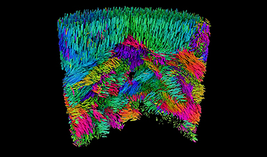
Their technique is known as X-ray linear dichroic orientation tomography, or XL-DOT for short. XL-DOT uses polarized X-rays from the Swiss Light Source SLS, to probe how materials absorb X-rays differently depending on the orientation of structural domains inside. By changing the polarization of the X-rays, while rotating the sample to capture images from different angles, the technique creates a three-dimensional map revealing the internal organization of the material.
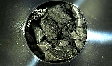
The team applied their method to a chunk of vanadium pentoxide catalyst about one micron in diameter, used in the production of sulfuric acid. Here, they could identify minute details in the catalyst`s structure including crystalline grains, boundaries where grains meet, and changes in the crystal orientation. They also identified topological defects in the catalyst. Such features directly affect the activity and stability of catalysts, so knowledge of this structure is crucial in optimizing performance.
Importantly, the method achieves high spatial resolution. Because X-rays have a short wavelength, the method can resolve structures just tens of nanometers in size, aligning with the sizes of features such as the crystalline grains.
"Linear dichroism has been used to measure anisotropies in materials for many years, but this is the first time it has been extended to 3D. We not only look inside, but with nanoscale resolution," says Valerio Scagnoli, Senior Scientist in the Mesoscopic Systems, a joint group between PSI and ETH Zurich. "This means that we now have access to information that was not previously visible, and we can achieve this in small but representative samples, several micrometers in size."
Leading the way with coherent X-rays
Although the researchers first had the idea for XL-DOT in 2019, it would take another five years to put it into practice. Together with complex experimental requirements, a major hurdle was extracting the three-dimensional map of crystal orientations from terabytes of raw data. This mathematical puzzle was overcome with the development of a dedicated reconstruction algorithm by Andreas Apseros, first author of the study, during his doctoral studies at PSI, funded by the Swiss National Science Foundation (SNSF).
The researchers believe that their success in developing XL-DOT is in part thanks to the long-term commitment to developing expertise with coherent X-rays at PSI, which led to unprecedented control and instrument stability at the coherent Small Angle X-ray Scattering (cSAXS) beamline: critical for the delicate measurements.
This is an area that is set to leap forward after the SLS 2.0 upgrade: "Coherence is where we're really set to gain with the upgrade," says Apseros. "We're looking at very weak signals, so with more coherent photons, we'll have more signal and can either go to more difficult materials or higher spatial resolution."
A way into the microstructure of diverse materials
Given the non-destructive nature of XL-DOT, the researchers foresee operando investigations of systems such as batteries as well as catalysts. "Catalyst bodies and cathode particles in batteries are typically between ten and fifty micrometers in size, so this is a reasonable next step," says Johannes Ihli, formerly of cSAXS and currently at the University of Oxford, who led the study.
Yet the new technique is not just useful for catalysts, the researchers emphasize. It is useful for all types of materials that exhibit ordered microstructures, whether biological tissues or advanced materials for information technology or energy storage.
Indeed, for the research team, the scientific motivation lies with probing the three-dimensional magnetic organization of materials. An example is the orientation of magnetic moments within antiferromagnetic materials. Here, the magnetic moments are aligned in alternating directions when going from atom to atom. Such materials maintain no net magnetization when measured at a distance, yet they do possess local order in the magnetic structure, a fact that is appealing for technological applications such as faster and more efficient data processing. "Our method is one of the only ways to probe this orientation," says Claire Donnelly, group leader Max Planck Institute for Chemical Physics of Solids in Dresden who, since carrying out her doctoral work in the Mesoscopic Systems group has maintained a strong collaboration with the team at PSI.
It was during this doctoral work that Donnelly together with the same team at PSI published in Nature a method to carry out magnetic tomography using circularly polarized X-rays (in contrast to XL-DOT, which uses linearly polarized X-rays). This has since been implemented in synchrotrons around the world.
With the groundwork for XL-DOT laid, the team hope that it will, in a similar way to its circularly polarized sibling, become a widely used technique at synchrotrons. Given the much wider range of samples that XL-DOT is relevant to and the importance of structural ordering to material performance, the impact of this latest method may be expected to be even greater. "Now that we've overcome many of the challenges, other beamlines can implement the technique. And we can help them to do it," adds Donnelly.
Reference: "X-ray Linear Dichroic Tomography of Crystallographic and Topological Defects" 11 December 2024, Nature.
News

RNA Recycling Extends Lifespan
Summary: Researchers discovered a biological “trash disposal” mechanism that directly controls how fast we age. While circular RNA has long been known to accumulate in cells as we get older, this study proves for the [...]

Cancer’s Deadly Paradox: How Tumors Break Their Own DNA To Keep Growing
Cancer’s strongest gene switches push DNA into damaging overdrive, creating repeated breaks and repairs that may fuel tumor evolution while exposing possible therapeutic weak spots. A new study indicates that cancer can harm its own genetic [...]

NanoMedical Brain/Cloud Interface – Explorations and Implications. A new book from Frank Boehm
New book from Frank Boehm, NanoappsMedical Inc Founder: This book explores the future hypothetical possibility that the cerebral cortex of the human brain might be seamlessly, safely, and securely connected with the Cloud via [...]

Our books now available worldwide!
Online Sellers other than Amazon, Routledge, and IOPP Indigo Global Health Care Equivalency in the Age of Nanotechnology, Nanomedicine and Artifcial Intelligence Global Health Care Equivalency In The Age Of Nanotechnology, Nanomedicine And Artificial [...]
Ryugu asteroid samples contain all DNA and RNA building blocks, bolstering origin-of-life theories
All the essential ingredients to make the DNA and RNA underpinning life on Earth have been discovered in samples collected from the asteroid Ryugu, scientists said Monday. The discovery comes after these building blocks [...]

Is Berberine Really a “Natural Ozempic”?
Often labeled a “natural Ozempic,” berberine is widely discussed as a metabolic aid. Yet research suggests its influence may lie deeper. In recent years, berberine has gained significant attention as a supposed “natural way” [...]
Viagra Ingredient Shows Promise for Rare Childhood Brain Disease in Surprising Study
A rare childhood disease with no approved treatment may have an unexpected new therapeutic candidate. Sildenafil, the active ingredient also sold under the brand name Viagra, may help reduce symptoms in people with Leigh [...]

In a first for China, Neuracle’s implantable brain-computer interface wins approval
In a landmark development, Neuracle Medical Technology has secured the country’s first-ever approval for an implantable brain-computer interface (BCI) system designed to restore hand motor function in patients with spinal cord injuries, in a [...]
A Cambridge Lab Mistake Reveals a Powerful New Way to Modify Drug Molecules
A surprising lab discovery reveals a light-powered way to tweak complex drugs faster, cleaner, and later in development. Researchers at the University of Cambridge have created a new technique for altering complex drug molecules [...]

New book from NanoappsMedical Inc – Molecular Manufacturing: The Future of Nanomedicine
This book explores the revolutionary potential of atomically precise manufacturing technologies to transform global healthcare, as well as practically every other sector across society. This forward-thinking volume examines how envisaged Factory@Home systems might enable the cost-effective [...]

Scientists Discover Simple Saliva Test That Reveals Hidden Diabetes Risk
Researchers have identified a potential new way to assess metabolic health using saliva instead of blood. High insulin levels in the blood, known as hyperinsulinemia, can reveal metabolic problems long before obvious symptoms appear. It is [...]

One Nasal Spray Could Protect Against COVID, Flu, Pneumonia, and More
A single nasal spray vaccine may one day protect against viruses, pneumonia, and even allergies. For decades, scientists have dreamed of creating a universal vaccine capable of protecting against many different pathogens. The idea [...]
New AI Model Predicts Cancer Spread With Incredible Accuracy
Scientists have developed an AI system that analyzes complex gene-expression signatures to estimate the likelihood that a tumor will spread. Why do some tumors spread throughout the body while others remain confined to their [...]

Scientists Discover DNA “Flips” That Supercharge Evolution
In Lake Malawi, hundreds of species of cichlid fish have evolved with astonishing speed, offering scientists a rare opportunity to study how biodiversity arises. Researchers have identified segments of “flipped” DNA that may allow fish to adapt rapidly [...]

Scientists Discover Why Some COVID Survivors Still Can’t Taste Food Years Later
A new study provides the first direct biological evidence explaining why some people continue to experience taste loss long after recovering from COVID-19. Researchers have uncovered specific biological changes in taste buds that could help [...]

Catching COVID significantly raises the risk of developing kidney disease, researchers find
Catching Covid significantly raises the risk of developing deadly kidney disease, research has shown. The virus was found to increase the chances that patients will develop the incurable condition by around 50 per cent. [...]