Images Description
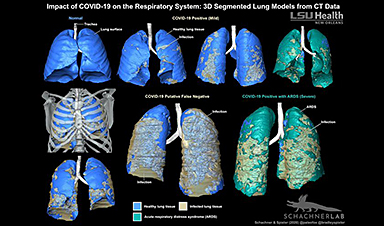
Three individuals were admitted to the hospital (ages 46–56; to men and one woman) with a multiday history of symptoms associated with the severe acute respiratory syndrome coronavirus 2 (SARS-CoV-2) and underwent contrast-enhanced thoracic CT due to worsening symptomatology. Three-dimensional (3D) digital models were created to visualise the extent of the disease within the respiratory system (figures 1 and 2) from the thin section (1 mm) data sets. All patients presented emergently with variable pulmonary symptoms ranging from mild to severe, including shortness of breath and all were febrile. Two of the patients were reverse transcription polymerase chain reaction (RT-PCR) positive for SARS-CoV-2 (figure 1B,D,F; figure 2C,D,G,H). The third patient was RT-PCR negative for SARS-CoV-2, but this was presumed to be a false-negative result given compelling clinical and imaging features indicative of COVID-19 (figures 1C,E and 2E,F). A fourth patient who presented to the emergency department and was suspected of having COVID-19 also underwent CT to assess for the possibility of pulmonary embolus (figures 1A and 2A,B). This individual tested negative for SARS-CoV-2, and the lungs were normal. All CT examinations were obtained using a Philips iCT 256 or iQon Spectral CT systems. Data were acquired using a 128×0.625 mm or 64×0.625 mm detector configuration with dual sampling, rotation time of 0.33 s (120 kVp 72 mAs).
Figure 1
Three-dimensional segmented surface models of normal, COVID-19 and suspected COVID-19 lungs in anterior view. (A) Healthy lung model of a 50-year-old man. (B) Lung model of a COVID-19-positive 46-year-old man with mild respiratory symptoms. (C) Lung model of a COVID-19-negative 56-year-old man with clinical suspicion for COVID-19. (D) Lung model of a COVID-19-positive 55-year-old woman with severe respiratory symptoms and ARDS. (E) C with a skeleton. (F) D but with the ARDS tissue made translucent to demonstrate the full extent of the ground glass opacities and consolidation throughout the parenchyma. ARDS, acute respiratory distress syndrome; C, consolidated infection; GGO, ground-glass opacities; HP, healthy parenchyma; PB, primary bronchus; TR, trachea. Colour key: blue, healthy tissue; yellow, consolidation and ground glass opacities; green, ARDS.
Figure 2
Three-dimensional (3D) segmented surface models of normal, COVID-19 and suspected COVID-19 lungs in posterior view (left) and coronal views with accompanying simplified diagrammatic illustrations of the coronal CTs demonstrating the infection sites. Healthy lungs of a 50-year-old man as a segmented 3D model in posterior view (A), a coronal contrast enhanced CT slice (B) and a diagrammatic illustration of B (C). COVID-19-positive 46-year-old man (mild respiratory symptoms) as a segmented 3D model in posterior view (D), a coronal contrast enhanced CT slice (E) and a diagrammatic illustration of E (F). Lungs of a COVID-19-negative 56-year-old man with clinical suspicion for COVID-19 as a segmented 3D model in posterior view (G), a coronal contrast-enhanced CT slice (H) and a diagrammatic illustration of H (I). Lungs of a COVID-19-positive 55-year-old woman with ARDS as a segmented 3D model in posterior view (J), a coronal contrast enhanced CT slice (K) and a diagrammatic illustration of K (L). Models demonstrate the relationship, distribution and full extent of the disease in 3D versus the single CT slice which only provides information on the localised position of the infection. ARDS, acute respiratory distress syndrome; C, consolidated infection; GGO, ground-glass opacities; HP, healthy parenchyma. Colour key: blue, healthy tissue; yellow, consolidation and ground glass opacities; green, ARDS. Images not to (relative) scale.
![]()
Image Credit: BMJ
News This Week

This Common Vitamin May Help Stop Prediabetes From Turning Into Diabetes
Vitamin D may help prevent type 2 diabetes in people with specific genetic variations, offering a possible path toward personalized diabetes prevention. More than 40% of U.S. adults have prediabetes, a condition in which [...]

Ebola, hantavirus: Is the world prepared for the next pandemic?
Funding cuts to health research and a growing antivaccine movement are making it harder than ever to respond to viruses. The World Health Organization (WHO) has declared that an Ebola outbreak in Uganda and [...]

May 2026 Healthcare News and Trends: Market Signals That Matter
Artificial intelligence is dominating headlines, telehealth has settled into a new normal, and digital health continues to promise transformation. However, much of what is being discussed in healthcare today reflects potential rather than reality. [...]
Scientists Rewire Donor Stem Cells To Outsmart Aggressive Blood Cancers
Researchers have tested a gene-edited stem cell transplant designed to shield healthy blood-forming cells from powerful cancer-targeting immunotherapies. For patients with highly aggressive blood cancers, stem cell transplantation can offer a rare chance at [...]

Recent Digital Health Trends, Insights and News – May 2026
Last month marked continued progress as digital health moves into its next phase — from AI expanding into drug discovery and core infrastructure to new federal pathways accelerating device access and home-based care. Together, [...]
Cancer Mystery Solved: Scientists Discover How Melanoma Becomes “Immortal”
Scientists have uncovered a previously overlooked mechanism that may help melanoma cells become effectively “immortal.” Cancer cells face a major problem before they can become deadly: They have to figure out how to stop [...]

How Visual Neurons Organize Thousands of Synaptic Inputs
Summary: A new study uncovered the organizational rules that determine how neurons in the primary visual cortex process information. By imaging both the cell bodies (soma) and the individual synapses (on dendritic spines) of [...]

Scientists Just Found a Surprising Way To Destroy “Forever Chemicals”
Scientists have uncovered a new mechanism that may help break down highly persistent PFAS pollutants. PFAS have earned the nickname “forever chemicals” for a reason. These industrial compounds are so chemically durable that they [...]

Scientists Discover Cheap Material That Kills Deadly Superbugs
A new sulfur-rich antimicrobial polymer shows strong effectiveness against fungal and bacterial pathogens and may offer an affordable solution to antimicrobial resistance. Antimicrobial resistance is creating growing challenges for both healthcare and food production, [...]

What to Know About Cicada, or BA.3.2, the Latest SARS-CoV-2 Variant Under Monitoring
Like periodical cicadas, the insects for which it is nicknamed, SARS-CoV-2 Omicron subvariant BA.3.2 is only just beginning to emerge after lying low for an extended period since it first appeared. Although it was [...]
Scientists Say This Simple Supplement May Actually Reverse Heart Disease
Scientists in Japan say a common supplement may actually help “unclog” certain diseased heart arteries from the inside out. A simple food supplement sold in Japan may have helped reverse a dangerous form of [...]

New breakthrough against radiation: Korean Scientists create revolutionary shield with nanotechnology
Korean Scientists develop new nanotechnology material capable of reducing radiation impacts in space missions, hospitals, and power plants. The search for more efficient protection technologies in extreme environments has just gained an important advance. Korean [...]
Scientists Just Discovered the Hidden Trick That Keeps Your Cells Alive
A strange bead-like motion inside cells may be the secret to keeping their DNA—and health—in balance. Mitochondria are often described as the power plants of the cell because they produce the energy cells need [...]
Scientists Discover Stem Cells That Could Regrow Teeth and Bone
Scientists just uncovered the cellular “blueprint” that could one day let us regrow real teeth. Researchers at Science Tokyo have uncovered two distinct stem cell lineages that play a central role in forming tooth [...]
Scientists Uncover Fatal Weakness in “Zombie Cells” Linked to Cancer
A newly identified weakness in “zombie” cells may open the door to more precise cancer treatments by turning their own survival strategy against them. A new class of drugs takes advantage of a recently [...]

Bowel and Ovarian Cancers Are Dramatically Rising in Young Adults, Scientists Aren’t Sure Why
Cancer incidence is increasing, especially among younger adults, and current risk factors don’t fully account for the trend. Scientists suggest other underlying causes may be contributing. Cancer patterns in England are shifting in a [...]