What if you could take a picture of every gene inside a living organism—not with light, but with DNA itself?
Scientists at the University of Chicago have pioneered a revolutionary imaging technique called volumetric DNA microscopy. It builds intricate 3D maps of genetic material by tagging and tracking molecular interactions, creating never-before-seen views inside organisms like zebrafish embryos.
New Window into Genetics
Traditional genetic sequencing can reveal a lot about the genetic material in a sample, such as a piece of tissue or a drop of blood, but it doesn’t show where specific genetic sequences are located within that sample, or how they relate to nearby genes and molecules.
To address this, researchers at the University of Chicago are developing a new technology that captures both the identity and location of genetic material. The method works by tagging individual DNA or RNA molecules and tracking how neighboring tags interact. These interactions are used to build a molecular network that reflects the spatial arrangement of genes, effectively creating a three-dimensional map of genetic activity. Known as volumetric DNA microscopy, the technique generates detailed 3D images of entire organisms from the inside out – down to the level of individual cells.

Imaging an Entire Organism
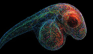
Joshua Weinstein, PhD, Assistant Professor of Medicine and Molecular Engineering at UChicago, has spent over a decade developing DNA microscopy, with support from the National Institutes of Health and the National Science Foundation. In a recent study published today (March 27) in Nature Biotechnology, Weinstein and postdoctoral researcher Nianchao Qian used the technique to produce a complete 3D DNA map of a zebrafish embryo—a widely used model for studying development and the nervous system.
“It’s a level of biology that no one has ever seen before,” Weinstein said. “To be able to see that kind of a view of nature from within a specimen is exhilarating.”
Rethinking Microscopy
Unlike traditional microscopes that use light or lenses, DNA microscopy creates images by calculating interactions among molecules, providing a new way to visualize genetic material in 3D. First, short DNA sequence tags called unique molecular identifiers (UMIs) are added to cells. They attach to DNA and RNA molecules and begin making copies of themselves. This starts a chemical reaction that creates new sequences, called unique event identifiers (UEIs), that are unique to each pairing.
It’s these pairings that help create the spatial map of where each genetic molecule is located. UMI pairs that are close together interact more frequently and generate more UEIs than those that are farther apart. Once the DNA and RNA are sequenced, a computational model reconstructs their original locations by analyzing the physical links between UMI-tags, creating a spatial map of gene expression.
Cell Phones and Cells: A Clever Analogy
Weinstein compares the technique to using data from cell phones pinging each other to determine people’s location in a city. Knowing the cell phone number or IP address of each person is like knowing the genetic sequence of one molecule, but if you can layer on their interactions with other phones nearby, you can work out their locations too.
“We can do this with cell phones and people, so why not do that with molecules and cells,” he said. “This turns the idea of imaging on its head. Rather than relying on an optical apparatus to shine light in, we can use biochemistry and DNA to form a massive network between molecules and encode their proximities to each other.”
Future Applications in Cancer and Immunotherapy
DNA microscopy doesn’t rely on prior knowledge of the genome or shape of a specimen, so it could be useful for understanding genetic expression in unique, unknown contexts. Tumors generate countless new genetic mutations, for example, so the tool would be able to map out the tumor microenvironment and where it interacts with the immune system. Immune cells interact with each other and respond to pathogens in context-specific ways, so DNA microscopy could help unravel those genetic mechanisms. Such applications could in turn guide more precise immunotherapy for cancer or tailor personalized vaccines.
“This is the critical foundation for being able to have truly comprehensive information about the ensemble of unique cells within the lymphatic system or tumor tissue,” Weinstein said. “There has still been this major gap in technology for allowing us to understand idiosyncratic tissue, and that’s what we’re trying to fill in here.”
DOI: 27 March 2025, Nature Biotechnology.
10.1038/s41587-025-02613-z
Additional funding for the study, “Spatial-transcriptomic imaging of an intact organism using volumetric DNA microscopy,” was provided by the Damon Runyon Foundation and the Moore Foundation.
News

This Deadly Disease Was Wiping Out Humans 5,500 Years Ago
A new study suggests plague was already a deadly threat 5,500 years ago, striking small hunter-gatherer communities long before cities and agriculture emerged. For centuries, plague has been remembered as the disease that devastated [...]

China closing in but US leads in biotech quality, commercial reach, survey finds
SAN DIEGO, June 22 (Reuters) - China, which now conducts more clinical drug trials, opens new tab than the U.S., still lags in the quality and commercial reach of its biomedical science, according to a recent survey, opens new [...]

New method generates renewable supply of progenitor immune cells
In a paper published in Cell, a USC Stem Cell-led team reports a new way of generating a renewable and expandable supply of the progenitor cells that give rise to macrophages. These immune cells help [...]

Scientists Just Discovered a Cellular Survival System That Was Never Supposed To Exist
A surprising backup pathway allows cells to make a crucial amino acid when their primary machinery fails. For decades, biologists believed cells had only one way to access a molecule they cannot live without. New [...]

Artificial cells gain porous membranes, enabling lab reactions and drug release
Artificial cells created in the laboratory offer a wide range of potential applications. Until now, however, their membranes—unlike those of real cells—have been virtually impermeable. Researchers at the Max Planck Institute for Polymer Research, [...]

Popular Weight-Loss Drugs Like Ozempic Linked to Lower Breast Cancer Risk
Ozempic and similar weight-loss drugs were linked to a striking 30% reduction in breast cancer risk in a study of more than 110,000 women. Popular weight-loss and diabetes medications such as Ozempic, Wegovy, Mounjaro, [...]

Stanford Scientists Discover Explosive New Type of Immune Cell
Scientists studying the remarkable regenerative abilities of planarian flatworms have uncovered a previously unknown type of immune cell with an unusually destructive defense strategy. What if an immune cell could wipe out nearby threats [...]

Big Pharma-backed SonoThera sounds off with $125M series B for bubble-based genetic delivery
Bay Area biotech SonoThera is bubbling to a clinical boil after raising a $125 million series B with the backing of some of the biggest names in pharma. Vida Ventures led the raise, with the venture [...]

Joint initiative of 5 EU countries calls for ‘unified approach’ to pharma framework amid US drug pricing pressure
With drug pricing pressure building from the U.S., a healthcare-focused consortium of five European countries is calling for a “unified approach” to strengthen Europe’s pharmaceutical framework and access to innovative medicines. Belgium, the Netherlands, [...]

Our books now available worldwide!
Online Sellers other than Amazon, Routledge, and IOPP Indigo Global Health Care Equivalency in the Age of Nanotechnology, Nanomedicine and Artifcial Intelligence Global Health Care Equivalency In The Age Of Nanotechnology, Nanomedicine And Artificial [...]

Molecular Manufacturing: The Future of Nanomedicine – New book from NanoappsMedical Inc.
This book explores the revolutionary potential of atomically precise manufacturing technologies to transform global healthcare, as well as practically every other sector across society. This forward-thinking volume examines how envisaged Factory@Home systems might enable the cost-effective [...]

NanoMedical Brain/Cloud Interface – Explorations and Implications. A new book from Frank Boehm
New book from Frank Boehm, NanoappsMedical Inc Founder: This book explores the future hypothetical possibility that the cerebral cortex of the human brain might be seamlessly, safely, and securely connected with the Cloud via [...]

New book from Nanoappsmedical Inc. – Global Health Care Equivalency
A new book by Frank Boehm, NanoappsMedical Inc. Founder. This groundbreaking volume explores the vision of a Global Health Care Equivalency (GHCE) system powered by artificial intelligence and quantum computing technologies, operating on secure [...]
UCLA Scientists Uncover a “Hidden Weakness” in Some of the World’s Deadliest Cancers
A new study has uncovered an unexpected vulnerability in some of the deadliest cancers. Researchers at UCLA have identified a previously hidden weakness in some of the most aggressive cancers, pointing to a possible new way [...]
AI-designed universal coronavirus vaccine clears first human trial
Key Takeaways Super-Antigen Technology: Uses AI and machine learning to analyze viral genomes, creating a single vaccine that targets essential features across entire virus families, including coronaviruses and Ebola. Human Trials & Safety: Phase [...]

Researchers Discover a Hidden Vitamin D Problem That Persists Year-Round
A new study suggests that some groups may not experience the expected seasonal boost in vitamin D levels, even during the sunniest months of the year. Many people assume that spending more time outdoors [...]
[/fusion_text][/fusion_builder_column][/fusion_builder_row][/fusion_builder_container]