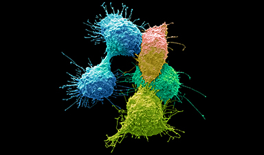
Researchers have integrated AI approaches from satellite mapping and community ecology to develop a tool to interpret data obtained from tumor tissue imaging, with the aim of implementing a more individualized approach to cancer care.
In recent years, huge advances have been made in tumor tissue imaging techniques. These developments have led to an incredibly rich amount of raw data. For instance, dozens of biological markers associated with the function and health of individual cells within tumor tissue can be quantified. This data contains information regarding the specific characteristics of different tumors, such as how they would respond to certain treatments. This could then be used to inform more effective and personalized treatment plans for individual cancer patients. However, strategies to effectively analyze and interpret this wealth of information have lagged behind these strides in imaging.
A collaboration between Karolinska Institutet and SciLifeLab (both Stockholm, Sweden) has led to the development of an AI tool capable of analyzing this information and converting it into useful outputs. This tool is referred to as niche-phenotype mapping (NIPMAP). Researchers integrated two unrelated approaches into NIPMAP. First, AI methods used to identify geographical features, such as conurbations and lakes, were used to map features on images of tumor tissues. Second, approaches used to identify how multiple species inhabit the same area, in a field called community ecology, were used to analyze the roles of individual cells and the relationships between them. This allowed for an intricate understanding of the position and role of individual cells within tumor tissues.
“We realized that the interpretation of tumor images is similar to the interpretation of satellite images and that the relationships between cells in a tissue are similar to the relationships between species in ecology,” elaborates Jean Hausser of the Karolinska Institutet, leader of this research.
The hope is that NIPMAP will be able to interpret the masses of data obtained from modern tumor tissue imaging techniques to identify key information such as which treatment options would be most effective.
Already, researchers have partnered with a cancer hospital in Lyon, France to attempt to use NIPMAP to reveal why only certain cancer patients respond to immunotherapy. A further collaboration with the Mayo Clinic in the USA aims to identify why certain breast cancer patients do not require chemotherapy. NIPMAP and other AI analysis techniques have the potential to drive a far greater degree of personalized and patient-tailored cancer treatment strategies through in-depth analysis of tumor features.
News
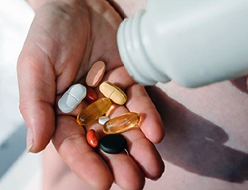
20-Year Study Finds Daily Multivitamins Don’t Extend Lifespan
A large, decades-long study of over 390,000 U.S. adults challenges a widespread assumption about daily multivitamins. Multivitamins are a daily habit for millions of Americans, often taken with the expectation that they will extend [...]
Novel Investment Paradigms for Regenerative Healthcare Ecosystems
Introduction The transition toward regenerative healthcare ecosystems—anchored in wellness optimization, disease prevention, eradication strategies, and healthy longevity—necessitates a structural reconfiguration of capital architectures, governance models, and incentive design. Regenerative healthcare, by definition, transcends episodic [...]
What If Consciousness Exists Beyond Your Brain
Scientists still don’t know how consciousness emerges from the brain. New ideas suggest it may not emerge at all, but instead be a basic feature of reality. Is consciousness produced by the brain, or [...]
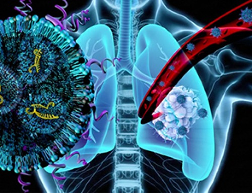
Scientists Discover Way To Treat Lung Cancer and Its Deadly Side Effect Together
A new approach using lipid nanoparticles to deliver genetic material is showing promise in tackling two major challenges in lung cancer at once.Researchers at Oregon State University have designed a new way to tackle two of [...]

Saunas Activate Your Immune System
A brief sauna session may quietly mobilize the immune system. A sauna session may do more than raise your heart rate and body temperature. A new study from Finland found that it also briefly [...]

Why music from your youth still has such an intense effect years later: A psychological perspective
You're driving, and suddenly a familiar song fills the air. Before you even know it, a wave of emotions comes over you – not just memories, but a deep, almost physical feeling. This powerful [...]
AI to antibody in days: breaking the wet lab bottleneck via high-throughput integration
The role of artificial intelligence (AI) in drug design has fundamentally shifted from a speculative tool to a central pillar of pharmaceutical research and development (R&D). Sino Biological plays a critical role in this [...]
Regenerative Healthcare by Design: Engineering Health-Centric Buildings and Urban Ecosystems
Introduction The next evolution of healthcare will not be confined to hospitals, clinics, or episodic interventions—it will be embedded into the infrastructure of everyday life. Regenerative health ecosystems require a systemic re-architecture of how [...]
Scientists Warn: Humanity Has Pushed the Planet Past Its Limits
Human population and consumption have surpassed Earth’s limits, increasing risks to climate and global stability. The Earth is already operating beyond its capacity to sustainably support the global population, according to new research highlighting [...]
Breakthrough Study Reveals Why Damaged Nerves Struggle To Heal
A newly identified molecular mechanism reveals how neurons weigh survival against repair after injury. Scientists at the Icahn School of Medicine at Mount Sinai have identified a molecular switch in neurons that limits the regrowth of [...]
Popular Vitamin B3 Supplements May Help Cancer Cells Survive, Scientists Warn
A new study raises important questions about widely used NAD+ supplements, suggesting that compounds often taken to boost energy and support healthy aging may have unintended consequences in cancer treatment. Millions of Americans take [...]
Scientists Discover Cancer Tumors Are “Addicted” to This Common Antioxidant
Cancer cells may be exploiting a common antioxidant as fuel, revealing a potential weakness that future therapies could target. Cancer cells may be tapping into an unexpected energy source: an antioxidant long associated with [...]
Nanotube injector transfers cytoplasmic contents and organelles between living cells safely
Cells are not isolated units; they continuously exchange proteins, genetic material, and even entire organelles with their neighbors. Intercellular transfer influences how tissues develop, respond to stress, and repair damage. In certain cancers, for [...]
CEO of America’s largest public hospital system is ready to replace radiologists with AI
The chief executive of America’s largest public hospital system says he is prepared to start replacing radiologists with artificial intelligence in some circumstances, once the regulatory landscape catches up. Mitchell H. Katz, MD, president [...]

Our books now available worldwide!
Online Sellers other than Amazon, Routledge, and IOPP Indigo Global Health Care Equivalency in the Age of Nanotechnology, Nanomedicine and Artifcial Intelligence Global Health Care Equivalency In The Age Of Nanotechnology, Nanomedicine And Artificial [...]

Study finds higher heart disease risk in long COVID patients
People with long COVID are at increased risk of developing cardiovascular disease, according to a new study from Karolinska Institutet published in eClinicalMedicine. The results show that the risk of conditions such as cardiac arrhythmias [...]